Magnesium »
PDB 6pek-6prv »
6pkz »
Magnesium in PDB 6pkz: Structure of Human Dna Polymerase Beta N279A Complexed with 8OA in the Template Base Paired with Incoming Non-Hydrolyzable Gtp
Enzymatic activity of Structure of Human Dna Polymerase Beta N279A Complexed with 8OA in the Template Base Paired with Incoming Non-Hydrolyzable Gtp
All present enzymatic activity of Structure of Human Dna Polymerase Beta N279A Complexed with 8OA in the Template Base Paired with Incoming Non-Hydrolyzable Gtp:
2.7.7.7;
2.7.7.7;
Protein crystallography data
The structure of Structure of Human Dna Polymerase Beta N279A Complexed with 8OA in the Template Base Paired with Incoming Non-Hydrolyzable Gtp, PDB code: 6pkz
was solved by
M.C.Koag,
S.Lee,
with X-Ray Crystallography technique. A brief refinement statistics is given in the table below:
| Resolution Low / High (Å) | 19.64 / 2.74 |
| Space group | P 1 21 1 |
| Cell size a, b, c (Å), α, β, γ (°) | 54.609, 78.555, 54.776, 90.00, 106.83, 90.00 |
| R / Rfree (%) | 17.5 / 24.9 |
Other elements in 6pkz:
The structure of Structure of Human Dna Polymerase Beta N279A Complexed with 8OA in the Template Base Paired with Incoming Non-Hydrolyzable Gtp also contains other interesting chemical elements:
| Sodium | (Na) | 2 atoms |
Magnesium Binding Sites:
The binding sites of Magnesium atom in the Structure of Human Dna Polymerase Beta N279A Complexed with 8OA in the Template Base Paired with Incoming Non-Hydrolyzable Gtp
(pdb code 6pkz). This binding sites where shown within
5.0 Angstroms radius around Magnesium atom.
In total only one binding site of Magnesium was determined in the Structure of Human Dna Polymerase Beta N279A Complexed with 8OA in the Template Base Paired with Incoming Non-Hydrolyzable Gtp, PDB code: 6pkz:
In total only one binding site of Magnesium was determined in the Structure of Human Dna Polymerase Beta N279A Complexed with 8OA in the Template Base Paired with Incoming Non-Hydrolyzable Gtp, PDB code: 6pkz:
Magnesium binding site 1 out of 1 in 6pkz
Go back to
Magnesium binding site 1 out
of 1 in the Structure of Human Dna Polymerase Beta N279A Complexed with 8OA in the Template Base Paired with Incoming Non-Hydrolyzable Gtp
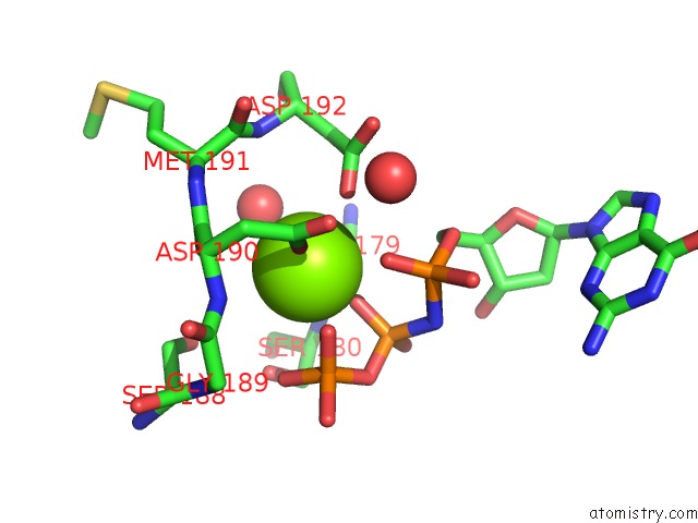
Mono view
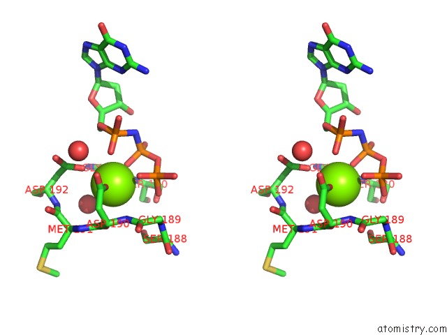
Stereo pair view
Mono view
Stereo pair view
A full contact list of Magnesium with other atoms in the Mg binding
site number 1 of Structure of Human Dna Polymerase Beta N279A Complexed with 8OA in the Template Base Paired with Incoming Non-Hydrolyzable Gtp within 5.0Å range:
|
Reference:
M.C.Koag,
S.Lee.
Structure of Human Dna Polymerase Beta N279A Complexed with 8OA in the Template Base Paired with Incoming Non-Hydrolyzable Gtp To Be Published.
Page generated: Tue Oct 1 14:11:29 2024
Last articles
Zn in 9JYWZn in 9IR4
Zn in 9IR3
Zn in 9GMX
Zn in 9GMW
Zn in 9JEJ
Zn in 9ERF
Zn in 9ERE
Zn in 9EGV
Zn in 9EGW