Magnesium »
PDB 6psq-6q5a »
6pz3 »
Magnesium in PDB 6pz3: Polymerase Eta-Catalyzed Insertion of Correct G Opposite Template Cytarabine (Arac) Residue
Enzymatic activity of Polymerase Eta-Catalyzed Insertion of Correct G Opposite Template Cytarabine (Arac) Residue
All present enzymatic activity of Polymerase Eta-Catalyzed Insertion of Correct G Opposite Template Cytarabine (Arac) Residue:
2.7.7.7;
2.7.7.7;
Protein crystallography data
The structure of Polymerase Eta-Catalyzed Insertion of Correct G Opposite Template Cytarabine (Arac) Residue, PDB code: 6pz3
was solved by
O.Rechkoblit,
A.K.Aggarwal,
with X-Ray Crystallography technique. A brief refinement statistics is given in the table below:
| Resolution Low / High (Å) | 42.53 / 2.40 |
| Space group | P 61 |
| Cell size a, b, c (Å), α, β, γ (°) | 98.221, 98.221, 81.348, 90.00, 90.00, 120.00 |
| R / Rfree (%) | 17.4 / 21.6 |
Magnesium Binding Sites:
The binding sites of Magnesium atom in the Polymerase Eta-Catalyzed Insertion of Correct G Opposite Template Cytarabine (Arac) Residue
(pdb code 6pz3). This binding sites where shown within
5.0 Angstroms radius around Magnesium atom.
In total 2 binding sites of Magnesium where determined in the Polymerase Eta-Catalyzed Insertion of Correct G Opposite Template Cytarabine (Arac) Residue, PDB code: 6pz3:
Jump to Magnesium binding site number: 1; 2;
In total 2 binding sites of Magnesium where determined in the Polymerase Eta-Catalyzed Insertion of Correct G Opposite Template Cytarabine (Arac) Residue, PDB code: 6pz3:
Jump to Magnesium binding site number: 1; 2;
Magnesium binding site 1 out of 2 in 6pz3
Go back to
Magnesium binding site 1 out
of 2 in the Polymerase Eta-Catalyzed Insertion of Correct G Opposite Template Cytarabine (Arac) Residue
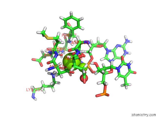
Mono view
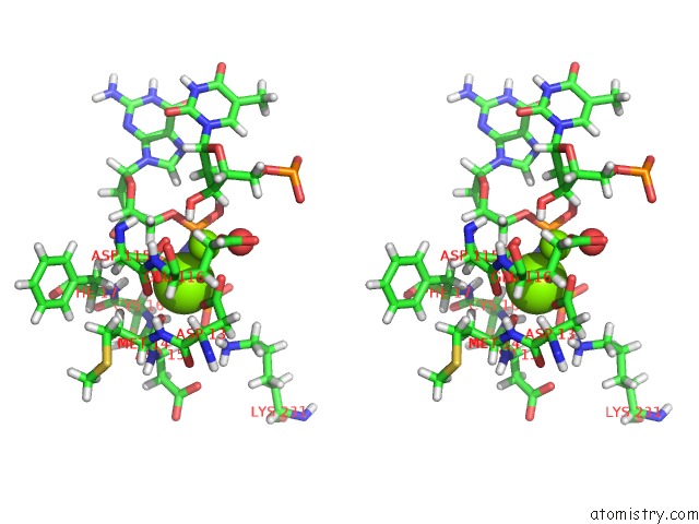
Stereo pair view
Mono view
Stereo pair view
A full contact list of Magnesium with other atoms in the Mg binding
site number 1 of Polymerase Eta-Catalyzed Insertion of Correct G Opposite Template Cytarabine (Arac) Residue within 5.0Å range:
|
Magnesium binding site 2 out of 2 in 6pz3
Go back to
Magnesium binding site 2 out
of 2 in the Polymerase Eta-Catalyzed Insertion of Correct G Opposite Template Cytarabine (Arac) Residue
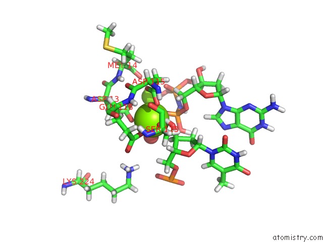
Mono view
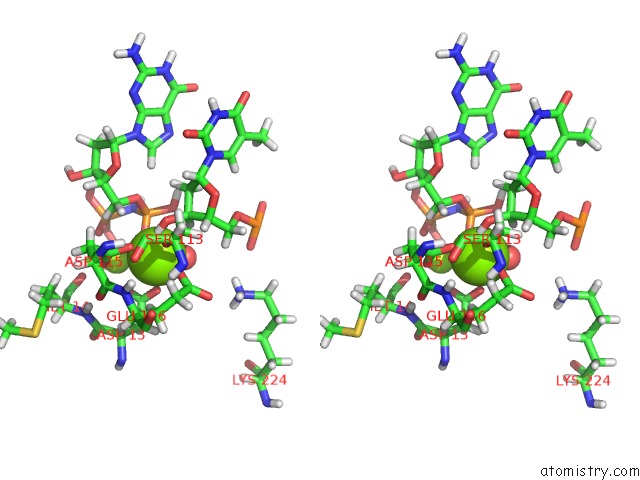
Stereo pair view
Mono view
Stereo pair view
A full contact list of Magnesium with other atoms in the Mg binding
site number 2 of Polymerase Eta-Catalyzed Insertion of Correct G Opposite Template Cytarabine (Arac) Residue within 5.0Å range:
|
Reference:
O.Rechkoblit,
R.E.Johnson,
A.Buku,
L.Prakash,
S.Prakash,
A.K.Aggarwal.
Structural Insights Into Mutagenicity of Anticancer Nucleoside Analog Cytarabine During Replication By Dna Polymerase Eta. Sci Rep V. 9 16400 2019.
ISSN: ESSN 2045-2322
PubMed: 31704958
DOI: 10.1038/S41598-019-52703-7
Page generated: Tue Oct 1 15:21:18 2024
ISSN: ESSN 2045-2322
PubMed: 31704958
DOI: 10.1038/S41598-019-52703-7
Last articles
Zn in 9J0NZn in 9J0O
Zn in 9J0P
Zn in 9FJX
Zn in 9EKB
Zn in 9C0F
Zn in 9CAH
Zn in 9CH0
Zn in 9CH3
Zn in 9CH1