Magnesium »
PDB 6q71-6qj6 »
6qii »
Magnesium in PDB 6qii: Xenon Derivatization of the F420-Reducing [Nife] Hydrogenase Complex From Methanosarcina Barkeri
Enzymatic activity of Xenon Derivatization of the F420-Reducing [Nife] Hydrogenase Complex From Methanosarcina Barkeri
All present enzymatic activity of Xenon Derivatization of the F420-Reducing [Nife] Hydrogenase Complex From Methanosarcina Barkeri:
1.12.98.1;
1.12.98.1;
Protein crystallography data
The structure of Xenon Derivatization of the F420-Reducing [Nife] Hydrogenase Complex From Methanosarcina Barkeri, PDB code: 6qii
was solved by
Y.Ilina,
C.Lorent,
S.Katz,
J.H.Jeoung,
S.Shima,
M.Horch,
I.Zebger,
H.Dobbek,
with X-Ray Crystallography technique. A brief refinement statistics is given in the table below:
| Resolution Low / High (Å) | 45.55 / 2.28 |
| Space group | F 2 3 |
| Cell size a, b, c (Å), α, β, γ (°) | 236.705, 236.705, 236.705, 90.00, 90.00, 90.00 |
| R / Rfree (%) | 17.6 / 22.7 |
Other elements in 6qii:
The structure of Xenon Derivatization of the F420-Reducing [Nife] Hydrogenase Complex From Methanosarcina Barkeri also contains other interesting chemical elements:
| Nickel | (Ni) | 1 atom |
| Xenon | (Xe) | 7 atoms |
| Iron | (Fe) | 19 atoms |
Magnesium Binding Sites:
The binding sites of Magnesium atom in the Xenon Derivatization of the F420-Reducing [Nife] Hydrogenase Complex From Methanosarcina Barkeri
(pdb code 6qii). This binding sites where shown within
5.0 Angstroms radius around Magnesium atom.
In total only one binding site of Magnesium was determined in the Xenon Derivatization of the F420-Reducing [Nife] Hydrogenase Complex From Methanosarcina Barkeri, PDB code: 6qii:
In total only one binding site of Magnesium was determined in the Xenon Derivatization of the F420-Reducing [Nife] Hydrogenase Complex From Methanosarcina Barkeri, PDB code: 6qii:
Magnesium binding site 1 out of 1 in 6qii
Go back to
Magnesium binding site 1 out
of 1 in the Xenon Derivatization of the F420-Reducing [Nife] Hydrogenase Complex From Methanosarcina Barkeri
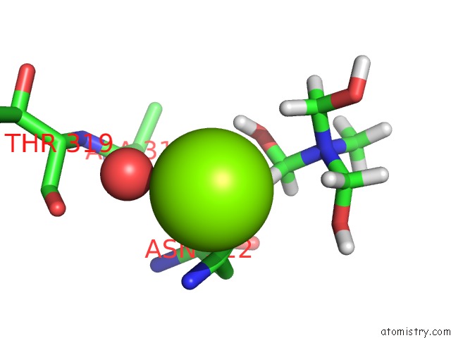
Mono view
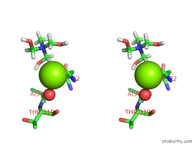
Stereo pair view
Mono view
Stereo pair view
A full contact list of Magnesium with other atoms in the Mg binding
site number 1 of Xenon Derivatization of the F420-Reducing [Nife] Hydrogenase Complex From Methanosarcina Barkeri within 5.0Å range:
|
Reference:
Y.Ilina,
C.Lorent,
S.Katz,
J.H.Jeoung,
S.Shima,
M.Horch,
I.Zebger,
H.Dobbek.
X-Ray Crystallography and Vibrational Spectroscopy Reveal the Key Determinants of Biocatalytic Dihydrogen Cycling By [Nife] Hydrogenases. Angew.Chem.Int.Ed.Engl. 2019.
ISSN: ESSN 1521-3773
PubMed: 31591784
DOI: 10.1002/ANIE.201908258
Page generated: Tue Oct 1 15:29:29 2024
ISSN: ESSN 1521-3773
PubMed: 31591784
DOI: 10.1002/ANIE.201908258
Last articles
Zn in 9MJ5Zn in 9HNW
Zn in 9G0L
Zn in 9FNE
Zn in 9DZN
Zn in 9E0I
Zn in 9D32
Zn in 9DAK
Zn in 8ZXC
Zn in 8ZUF