Magnesium »
PDB 6quy-6r5k »
6r1b »
Magnesium in PDB 6r1b: Crystal Structure of Ugpb From Mycobacterium Tuberculosis in Complex with Glycerophosphocholine
Protein crystallography data
The structure of Crystal Structure of Ugpb From Mycobacterium Tuberculosis in Complex with Glycerophosphocholine, PDB code: 6r1b
was solved by
J.Fenn,
R.Nepravishta,
C.S.Guy,
J.Harrison,
J.Angulo,
A.D.Cameron,
E.Fullam,
with X-Ray Crystallography technique. A brief refinement statistics is given in the table below:
| Resolution Low / High (Å) | 38.69 / 2.27 |
| Space group | P 21 21 21 |
| Cell size a, b, c (Å), α, β, γ (°) | 46.110, 169.860, 213.310, 90.00, 90.00, 90.00 |
| R / Rfree (%) | 20.6 / 25.6 |
Magnesium Binding Sites:
The binding sites of Magnesium atom in the Crystal Structure of Ugpb From Mycobacterium Tuberculosis in Complex with Glycerophosphocholine
(pdb code 6r1b). This binding sites where shown within
5.0 Angstroms radius around Magnesium atom.
In total 5 binding sites of Magnesium where determined in the Crystal Structure of Ugpb From Mycobacterium Tuberculosis in Complex with Glycerophosphocholine, PDB code: 6r1b:
Jump to Magnesium binding site number: 1; 2; 3; 4; 5;
In total 5 binding sites of Magnesium where determined in the Crystal Structure of Ugpb From Mycobacterium Tuberculosis in Complex with Glycerophosphocholine, PDB code: 6r1b:
Jump to Magnesium binding site number: 1; 2; 3; 4; 5;
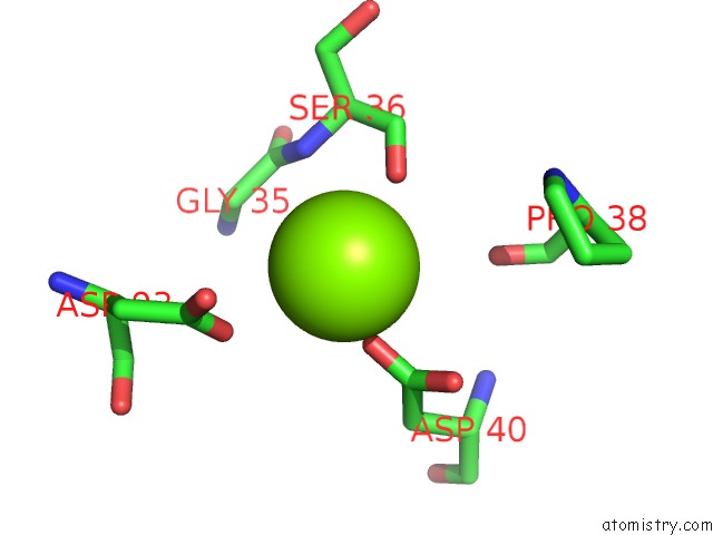
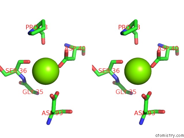
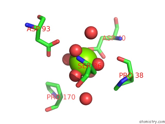
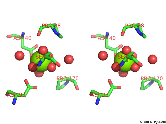
Magnesium binding site 1 out of 5 in 6r1b
Go back to
Magnesium binding site 1 out
of 5 in the Crystal Structure of Ugpb From Mycobacterium Tuberculosis in Complex with Glycerophosphocholine
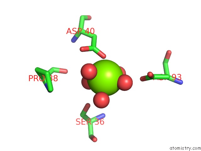
Mono view
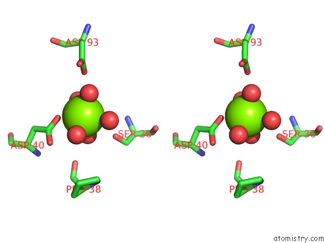
Stereo pair view
Mono view
Stereo pair view
A full contact list of Magnesium with other atoms in the Mg binding
site number 1 of Crystal Structure of Ugpb From Mycobacterium Tuberculosis in Complex with Glycerophosphocholine within 5.0Å range:
|
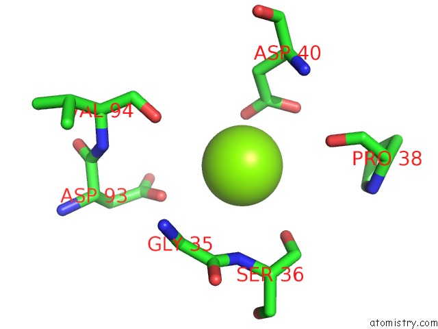
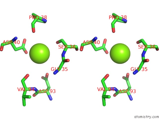
Magnesium binding site 2 out of 5 in 6r1b
Go back to
Magnesium binding site 2 out
of 5 in the Crystal Structure of Ugpb From Mycobacterium Tuberculosis in Complex with Glycerophosphocholine
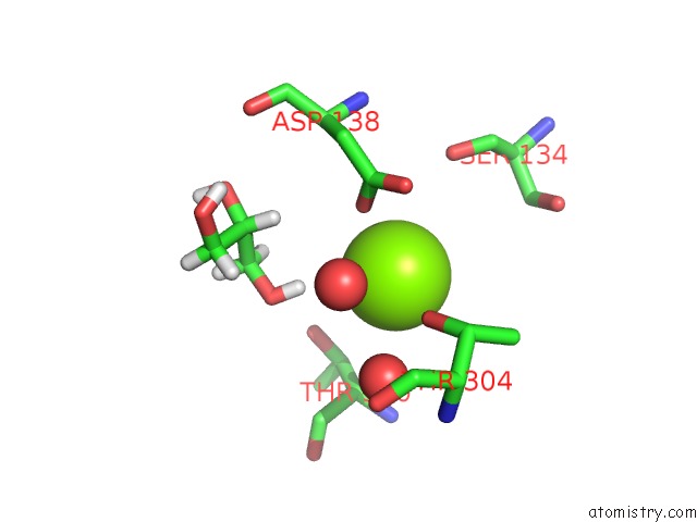
Mono view
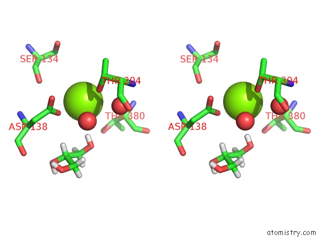
Stereo pair view
Mono view
Stereo pair view
A full contact list of Magnesium with other atoms in the Mg binding
site number 2 of Crystal Structure of Ugpb From Mycobacterium Tuberculosis in Complex with Glycerophosphocholine within 5.0Å range:
|
Magnesium binding site 3 out of 5 in 6r1b
Go back to
Magnesium binding site 3 out
of 5 in the Crystal Structure of Ugpb From Mycobacterium Tuberculosis in Complex with Glycerophosphocholine
Mono view
Stereo pair view
Mono view
Stereo pair view
A full contact list of Magnesium with other atoms in the Mg binding
site number 3 of Crystal Structure of Ugpb From Mycobacterium Tuberculosis in Complex with Glycerophosphocholine within 5.0Å range:
|
Magnesium binding site 4 out of 5 in 6r1b
Go back to
Magnesium binding site 4 out
of 5 in the Crystal Structure of Ugpb From Mycobacterium Tuberculosis in Complex with Glycerophosphocholine
Mono view
Stereo pair view
Mono view
Stereo pair view
A full contact list of Magnesium with other atoms in the Mg binding
site number 4 of Crystal Structure of Ugpb From Mycobacterium Tuberculosis in Complex with Glycerophosphocholine within 5.0Å range:
|
Magnesium binding site 5 out of 5 in 6r1b
Go back to
Magnesium binding site 5 out
of 5 in the Crystal Structure of Ugpb From Mycobacterium Tuberculosis in Complex with Glycerophosphocholine
Mono view
Stereo pair view
Mono view
Stereo pair view
A full contact list of Magnesium with other atoms in the Mg binding
site number 5 of Crystal Structure of Ugpb From Mycobacterium Tuberculosis in Complex with Glycerophosphocholine within 5.0Å range:
|
Reference:
J.S.Fenn,
R.Nepravishta,
C.S.Guy,
J.Harrison,
J.Angulo,
A.D.Cameron,
E.Fullam.
Structural Basis of Glycerophosphodiester Recognition By Themycobacterium Tuberculosissubstrate-Binding Protein Ugpb. Acs Chem.Biol. V. 14 1879 2019.
ISSN: ESSN 1554-8937
PubMed: 31433162
DOI: 10.1021/ACSCHEMBIO.9B00204
Page generated: Tue Oct 1 16:29:48 2024
ISSN: ESSN 1554-8937
PubMed: 31433162
DOI: 10.1021/ACSCHEMBIO.9B00204
Last articles
Zn in 9J0NZn in 9J0O
Zn in 9J0P
Zn in 9FJX
Zn in 9EKB
Zn in 9C0F
Zn in 9CAH
Zn in 9CH0
Zn in 9CH3
Zn in 9CH1