Magnesium »
PDB 6quy-6r5k »
6r2d »
Magnesium in PDB 6r2d: Crystal Structure of the Suca Domain of Mycobacterium Smegmatis Kgd After Soaking with Succinylphosphonate Phosphonoethyl Ester, Followed By Temperature Increase
Enzymatic activity of Crystal Structure of the Suca Domain of Mycobacterium Smegmatis Kgd After Soaking with Succinylphosphonate Phosphonoethyl Ester, Followed By Temperature Increase
All present enzymatic activity of Crystal Structure of the Suca Domain of Mycobacterium Smegmatis Kgd After Soaking with Succinylphosphonate Phosphonoethyl Ester, Followed By Temperature Increase:
1.2.4.2; 2.2.1.5; 2.3.1.61; 4.1.1.71;
1.2.4.2; 2.2.1.5; 2.3.1.61; 4.1.1.71;
Protein crystallography data
The structure of Crystal Structure of the Suca Domain of Mycobacterium Smegmatis Kgd After Soaking with Succinylphosphonate Phosphonoethyl Ester, Followed By Temperature Increase, PDB code: 6r2d
was solved by
T.Wagner,
P.M.Alzari,
M.Bellinzoni,
with X-Ray Crystallography technique. A brief refinement statistics is given in the table below:
| Resolution Low / High (Å) | 33.44 / 2.30 |
| Space group | P 1 |
| Cell size a, b, c (Å), α, β, γ (°) | 80.840, 83.730, 160.570, 99.52, 98.94, 100.81 |
| R / Rfree (%) | 21.7 / 24.2 |
Other elements in 6r2d:
The structure of Crystal Structure of the Suca Domain of Mycobacterium Smegmatis Kgd After Soaking with Succinylphosphonate Phosphonoethyl Ester, Followed By Temperature Increase also contains other interesting chemical elements:
| Calcium | (Ca) | 4 atoms |
Magnesium Binding Sites:
The binding sites of Magnesium atom in the Crystal Structure of the Suca Domain of Mycobacterium Smegmatis Kgd After Soaking with Succinylphosphonate Phosphonoethyl Ester, Followed By Temperature Increase
(pdb code 6r2d). This binding sites where shown within
5.0 Angstroms radius around Magnesium atom.
In total 4 binding sites of Magnesium where determined in the Crystal Structure of the Suca Domain of Mycobacterium Smegmatis Kgd After Soaking with Succinylphosphonate Phosphonoethyl Ester, Followed By Temperature Increase, PDB code: 6r2d:
Jump to Magnesium binding site number: 1; 2; 3; 4;
In total 4 binding sites of Magnesium where determined in the Crystal Structure of the Suca Domain of Mycobacterium Smegmatis Kgd After Soaking with Succinylphosphonate Phosphonoethyl Ester, Followed By Temperature Increase, PDB code: 6r2d:
Jump to Magnesium binding site number: 1; 2; 3; 4;
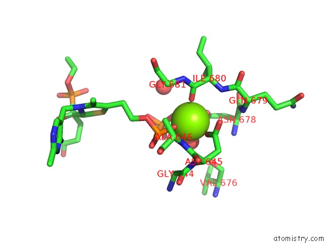
Magnesium binding site 1 out of 4 in 6r2d
Go back to
Magnesium binding site 1 out
of 4 in the Crystal Structure of the Suca Domain of Mycobacterium Smegmatis Kgd After Soaking with Succinylphosphonate Phosphonoethyl Ester, Followed By Temperature Increase
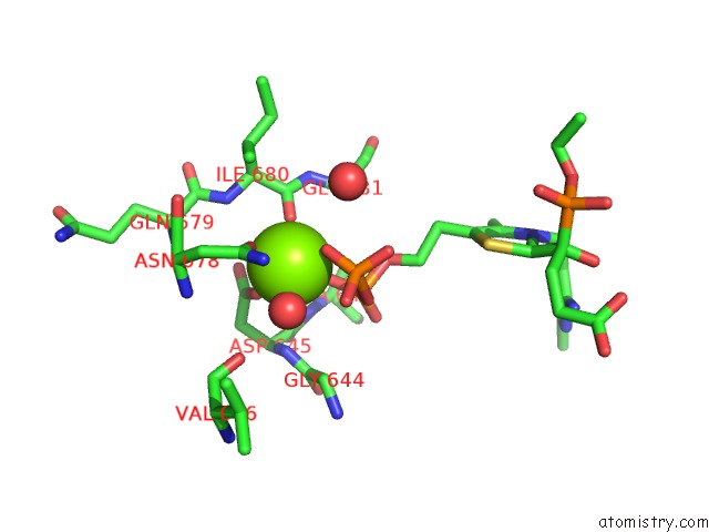
Mono view
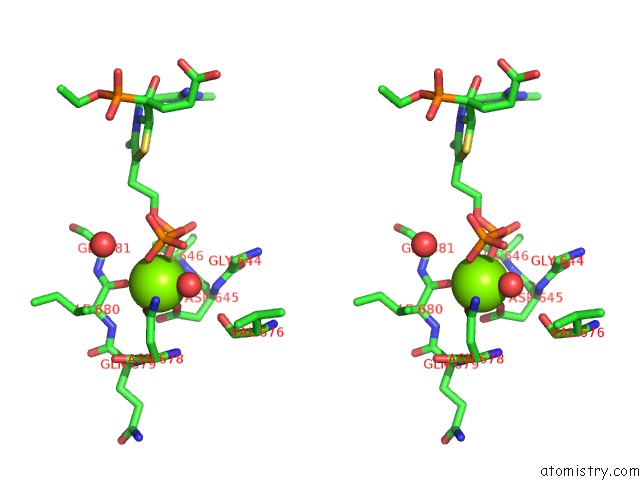
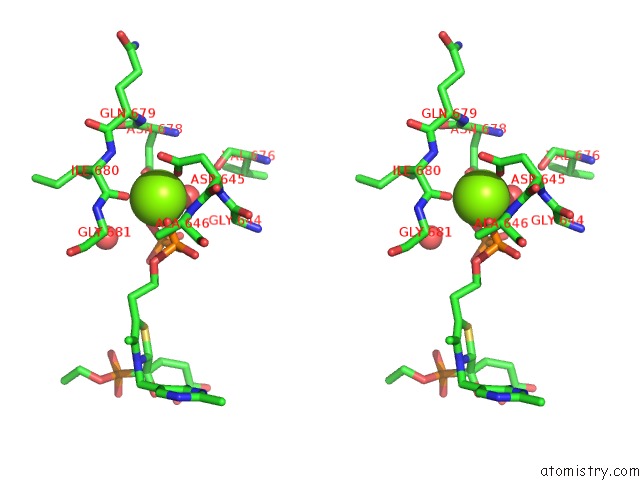
Stereo pair view
Mono view
Stereo pair view
A full contact list of Magnesium with other atoms in the Mg binding
site number 1 of Crystal Structure of the Suca Domain of Mycobacterium Smegmatis Kgd After Soaking with Succinylphosphonate Phosphonoethyl Ester, Followed By Temperature Increase within 5.0Å range:
|
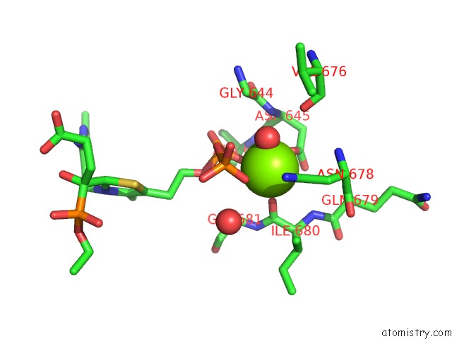
Magnesium binding site 2 out of 4 in 6r2d
Go back to
Magnesium binding site 2 out
of 4 in the Crystal Structure of the Suca Domain of Mycobacterium Smegmatis Kgd After Soaking with Succinylphosphonate Phosphonoethyl Ester, Followed By Temperature Increase
Mono view
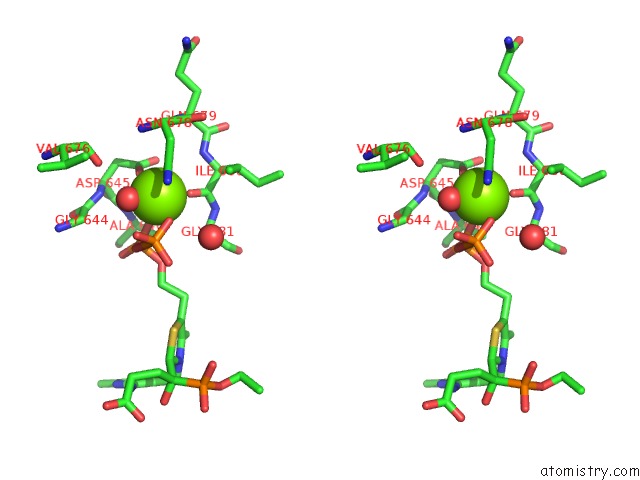
Stereo pair view
Mono view
Stereo pair view
A full contact list of Magnesium with other atoms in the Mg binding
site number 2 of Crystal Structure of the Suca Domain of Mycobacterium Smegmatis Kgd After Soaking with Succinylphosphonate Phosphonoethyl Ester, Followed By Temperature Increase within 5.0Å range:
|
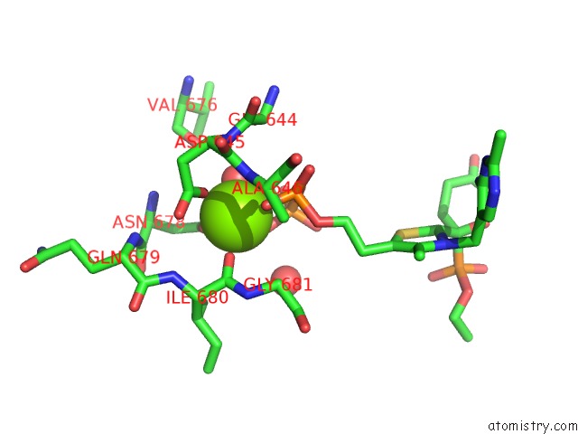
Magnesium binding site 3 out of 4 in 6r2d
Go back to
Magnesium binding site 3 out
of 4 in the Crystal Structure of the Suca Domain of Mycobacterium Smegmatis Kgd After Soaking with Succinylphosphonate Phosphonoethyl Ester, Followed By Temperature Increase
Mono view
Stereo pair view
Mono view
Stereo pair view
A full contact list of Magnesium with other atoms in the Mg binding
site number 3 of Crystal Structure of the Suca Domain of Mycobacterium Smegmatis Kgd After Soaking with Succinylphosphonate Phosphonoethyl Ester, Followed By Temperature Increase within 5.0Å range:
|
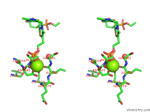
Magnesium binding site 4 out of 4 in 6r2d
Go back to
Magnesium binding site 4 out
of 4 in the Crystal Structure of the Suca Domain of Mycobacterium Smegmatis Kgd After Soaking with Succinylphosphonate Phosphonoethyl Ester, Followed By Temperature Increase
Mono view
Stereo pair view
Mono view
Stereo pair view
A full contact list of Magnesium with other atoms in the Mg binding
site number 4 of Crystal Structure of the Suca Domain of Mycobacterium Smegmatis Kgd After Soaking with Succinylphosphonate Phosphonoethyl Ester, Followed By Temperature Increase within 5.0Å range:
|
Reference:
T.Wagner,
A.Boyko,
P.M.Alzari,
V.I.Bunik,
M.Bellinzoni.
Conformational Transitions in the Active Site of Mycobacterial 2-Oxoglutarate Dehydrogenase Upon Binding Phosphonate Analogues of 2-Oxoglutarate: From A Michaelis-Like Complex to Thdp Adducts. J.Struct.Biol. V. 208 182 2019.
ISSN: ESSN 1095-8657
PubMed: 31476368
DOI: 10.1016/J.JSB.2019.08.012
Page generated: Tue Oct 1 16:30:34 2024
ISSN: ESSN 1095-8657
PubMed: 31476368
DOI: 10.1016/J.JSB.2019.08.012
Last articles
Zn in 9J0NZn in 9J0O
Zn in 9J0P
Zn in 9FJX
Zn in 9EKB
Zn in 9C0F
Zn in 9CAH
Zn in 9CH0
Zn in 9CH3
Zn in 9CH1