Magnesium »
PDB 6quz-6r5l »
6r5i »
Magnesium in PDB 6r5i: The Crystal Structure of the Glycoside Hydrolase Bglx From P. Aeruginosa
Protein crystallography data
The structure of The Crystal Structure of the Glycoside Hydrolase Bglx From P. Aeruginosa, PDB code: 6r5i
was solved by
M.T.Batuecas,
J.A.Hermoso,
with X-Ray Crystallography technique. A brief refinement statistics is given in the table below:
| Resolution Low / High (Å) | 19.93 / 1.80 |
| Space group | P 1 |
| Cell size a, b, c (Å), α, β, γ (°) | 71.079, 73.701, 81.534, 65.58, 73.91, 69.70 |
| R / Rfree (%) | 16.8 / 20.7 |
Magnesium Binding Sites:
The binding sites of Magnesium atom in the The Crystal Structure of the Glycoside Hydrolase Bglx From P. Aeruginosa
(pdb code 6r5i). This binding sites where shown within
5.0 Angstroms radius around Magnesium atom.
In total 2 binding sites of Magnesium where determined in the The Crystal Structure of the Glycoside Hydrolase Bglx From P. Aeruginosa, PDB code: 6r5i:
Jump to Magnesium binding site number: 1; 2;
In total 2 binding sites of Magnesium where determined in the The Crystal Structure of the Glycoside Hydrolase Bglx From P. Aeruginosa, PDB code: 6r5i:
Jump to Magnesium binding site number: 1; 2;
Magnesium binding site 1 out of 2 in 6r5i
Go back to
Magnesium binding site 1 out
of 2 in the The Crystal Structure of the Glycoside Hydrolase Bglx From P. Aeruginosa
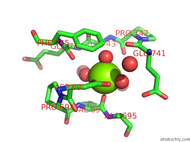
Mono view
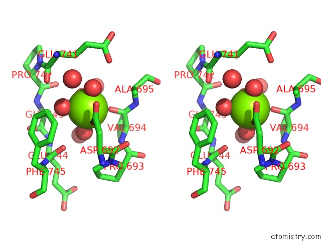
Stereo pair view
Mono view
Stereo pair view
A full contact list of Magnesium with other atoms in the Mg binding
site number 1 of The Crystal Structure of the Glycoside Hydrolase Bglx From P. Aeruginosa within 5.0Å range:
|
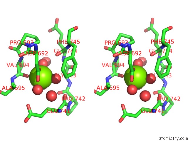
Magnesium binding site 2 out of 2 in 6r5i
Go back to
Magnesium binding site 2 out
of 2 in the The Crystal Structure of the Glycoside Hydrolase Bglx From P. Aeruginosa
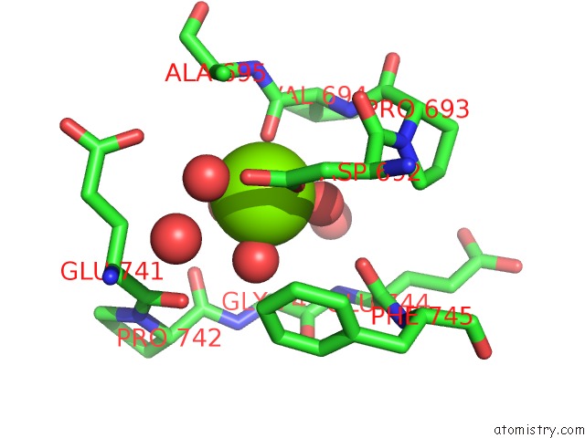
Mono view
Stereo pair view
Mono view
Stereo pair view
A full contact list of Magnesium with other atoms in the Mg binding
site number 2 of The Crystal Structure of the Glycoside Hydrolase Bglx From P. Aeruginosa within 5.0Å range:
|
Reference:
K.V.Mahasenan,
M.T.Batuecas,
S.De Benedetti,
C.Kim,
N.Rana,
M.Lee,
D.Hesek,
J.F.Fisher,
J.Sanz-Aparicio,
J.A.Hermoso,
S.Mobashery.
Catalytic Cycle of Glycoside Hydrolase Bglx Frompseudomonas Aeruginosaand Its Implications For Biofilm Formation. Acs Chem.Biol. V. 15 189 2020.
ISSN: ESSN 1554-8937
PubMed: 31877028
DOI: 10.1021/ACSCHEMBIO.9B00754
Page generated: Tue Oct 1 16:34:31 2024
ISSN: ESSN 1554-8937
PubMed: 31877028
DOI: 10.1021/ACSCHEMBIO.9B00754
Last articles
Ca in 5V2CCa in 5V4D
Ca in 5V6M
Ca in 5UZ8
Ca in 5V1N
Ca in 5V1F
Ca in 5V0S
Ca in 5V0Q
Ca in 5V03
Ca in 5UWN