Magnesium »
PDB 6rl3-6rux »
6rr6 »
Magnesium in PDB 6rr6: Structure of 100% Reduced Kpdyp
Protein crystallography data
The structure of Structure of 100% Reduced Kpdyp, PDB code: 6rr6
was solved by
V.Pfanzagl,
J.Beale,
S.Hofbauer,
with X-Ray Crystallography technique. A brief refinement statistics is given in the table below:
| Resolution Low / High (Å) | 47.67 / 1.90 |
| Space group | P 1 21 1 |
| Cell size a, b, c (Å), α, β, γ (°) | 50.889, 76.647, 76.579, 90.00, 108.01, 90.00 |
| R / Rfree (%) | 18.1 / 19.9 |
Other elements in 6rr6:
The structure of Structure of 100% Reduced Kpdyp also contains other interesting chemical elements:
| Iron | (Fe) | 2 atoms |
Magnesium Binding Sites:
The binding sites of Magnesium atom in the Structure of 100% Reduced Kpdyp
(pdb code 6rr6). This binding sites where shown within
5.0 Angstroms radius around Magnesium atom.
In total 2 binding sites of Magnesium where determined in the Structure of 100% Reduced Kpdyp, PDB code: 6rr6:
Jump to Magnesium binding site number: 1; 2;
In total 2 binding sites of Magnesium where determined in the Structure of 100% Reduced Kpdyp, PDB code: 6rr6:
Jump to Magnesium binding site number: 1; 2;
Magnesium binding site 1 out of 2 in 6rr6
Go back to
Magnesium binding site 1 out
of 2 in the Structure of 100% Reduced Kpdyp
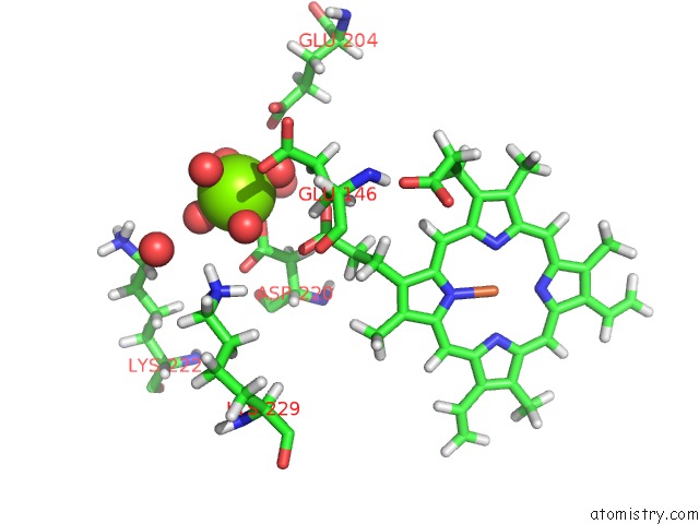
Mono view
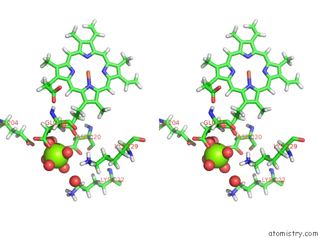
Stereo pair view
Mono view
Stereo pair view
A full contact list of Magnesium with other atoms in the Mg binding
site number 1 of Structure of 100% Reduced Kpdyp within 5.0Å range:
|
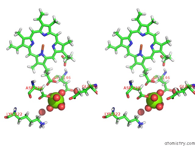
Magnesium binding site 2 out of 2 in 6rr6
Go back to
Magnesium binding site 2 out
of 2 in the Structure of 100% Reduced Kpdyp
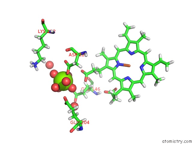
Mono view
Stereo pair view
Mono view
Stereo pair view
A full contact list of Magnesium with other atoms in the Mg binding
site number 2 of Structure of 100% Reduced Kpdyp within 5.0Å range:
|
Reference:
V.Pfanzagl,
J.H.Beale,
H.Michlits,
D.Schmidt,
T.Gabler,
C.Obinger,
K.Djinovic-Carugo,
S.Hofbauer.
X-Ray-Induced Photoreduction of Heme Metal Centers Rapidly Induces Active-Site Perturbations in A Protein-Independent Manner. J.Biol.Chem. V. 295 13488 2020.
ISSN: ESSN 1083-351X
PubMed: 32723869
DOI: 10.1074/JBC.RA120.014087
Page generated: Tue Oct 1 17:31:03 2024
ISSN: ESSN 1083-351X
PubMed: 32723869
DOI: 10.1074/JBC.RA120.014087
Last articles
Zn in 9J0NZn in 9J0O
Zn in 9J0P
Zn in 9FJX
Zn in 9EKB
Zn in 9C0F
Zn in 9CAH
Zn in 9CH0
Zn in 9CH3
Zn in 9CH1