Magnesium »
PDB 6rl3-6rux »
6rsw »
Magnesium in PDB 6rsw: Hfd Domain of Mouse CAP1 Bound to the Pointed End of G-Actin
Protein crystallography data
The structure of Hfd Domain of Mouse CAP1 Bound to the Pointed End of G-Actin, PDB code: 6rsw
was solved by
T.Kotila,
K.Kogan,
P.Lappalainen,
with X-Ray Crystallography technique. A brief refinement statistics is given in the table below:
| Resolution Low / High (Å) | 41.45 / 1.95 |
| Space group | P 1 21 1 |
| Cell size a, b, c (Å), α, β, γ (°) | 87.370, 54.490, 87.830, 90.00, 93.61, 90.00 |
| R / Rfree (%) | 16.6 / 19.4 |
Magnesium Binding Sites:
The binding sites of Magnesium atom in the Hfd Domain of Mouse CAP1 Bound to the Pointed End of G-Actin
(pdb code 6rsw). This binding sites where shown within
5.0 Angstroms radius around Magnesium atom.
In total 2 binding sites of Magnesium where determined in the Hfd Domain of Mouse CAP1 Bound to the Pointed End of G-Actin, PDB code: 6rsw:
Jump to Magnesium binding site number: 1; 2;
In total 2 binding sites of Magnesium where determined in the Hfd Domain of Mouse CAP1 Bound to the Pointed End of G-Actin, PDB code: 6rsw:
Jump to Magnesium binding site number: 1; 2;
Magnesium binding site 1 out of 2 in 6rsw
Go back to
Magnesium binding site 1 out
of 2 in the Hfd Domain of Mouse CAP1 Bound to the Pointed End of G-Actin
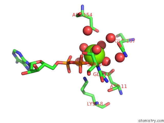
Mono view
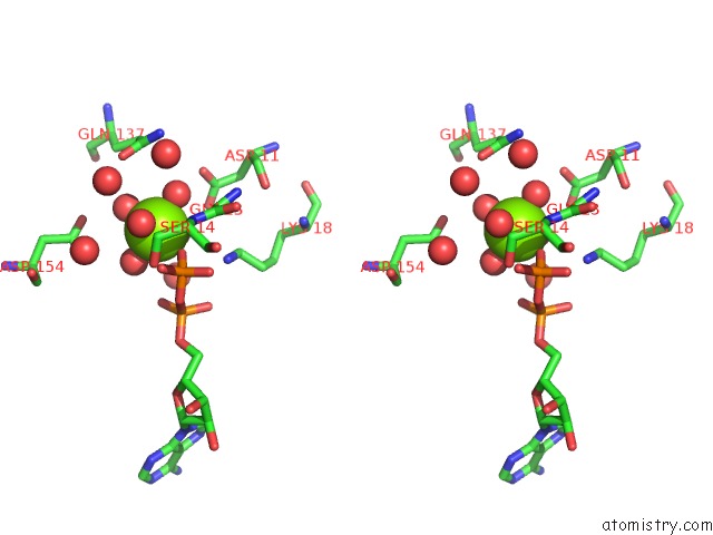
Stereo pair view
Mono view
Stereo pair view
A full contact list of Magnesium with other atoms in the Mg binding
site number 1 of Hfd Domain of Mouse CAP1 Bound to the Pointed End of G-Actin within 5.0Å range:
|
Magnesium binding site 2 out of 2 in 6rsw
Go back to
Magnesium binding site 2 out
of 2 in the Hfd Domain of Mouse CAP1 Bound to the Pointed End of G-Actin
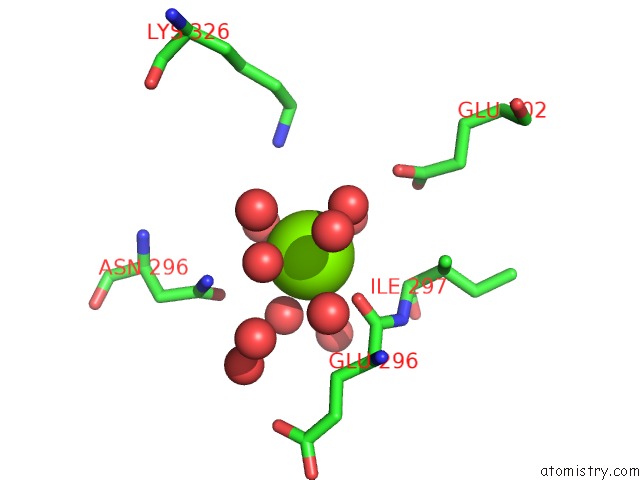
Mono view
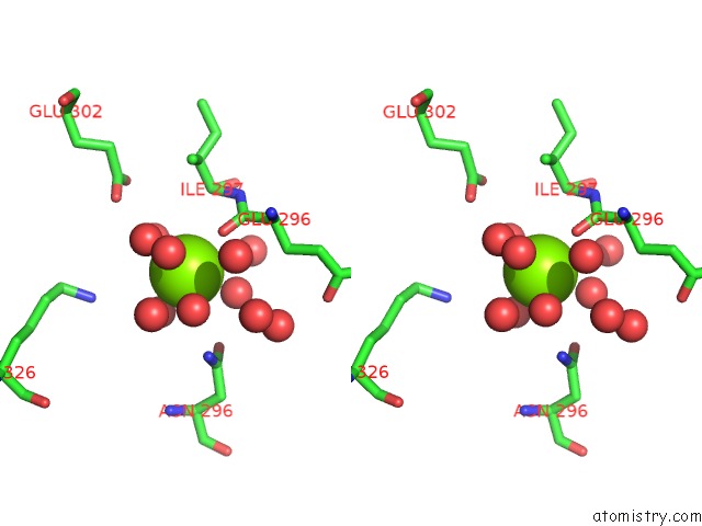
Stereo pair view
Mono view
Stereo pair view
A full contact list of Magnesium with other atoms in the Mg binding
site number 2 of Hfd Domain of Mouse CAP1 Bound to the Pointed End of G-Actin within 5.0Å range:
|
Reference:
T.Kotila,
H.Wioland,
G.Enkavi,
K.Kogan,
I.Vattulainen,
A.Jegou,
G.Romet-Lemonne,
P.Lappalainen.
Mechanism of Synergistic Actin Filament Pointed End Depolymerization By Cyclase-Associated Protein and Cofilin. Nat Commun V. 10 5320 2019.
ISSN: ESSN 2041-1723
PubMed: 31757941
DOI: 10.1038/S41467-019-13213-2
Page generated: Tue Oct 1 17:31:12 2024
ISSN: ESSN 2041-1723
PubMed: 31757941
DOI: 10.1038/S41467-019-13213-2
Last articles
Zn in 9J0NZn in 9J0O
Zn in 9J0P
Zn in 9FJX
Zn in 9EKB
Zn in 9C0F
Zn in 9CAH
Zn in 9CH0
Zn in 9CH3
Zn in 9CH1