Magnesium »
PDB 6rla-6rw0 »
6rux »
Magnesium in PDB 6rux: P46, An Immunodominant Surface Protein From Mycoplasma Hyopneumoniae
Protein crystallography data
The structure of P46, An Immunodominant Surface Protein From Mycoplasma Hyopneumoniae, PDB code: 6rux
was solved by
A.Guasch,
I.Fita,
with X-Ray Crystallography technique. A brief refinement statistics is given in the table below:
| Resolution Low / High (Å) | 60.00 / 2.50 |
| Space group | P 31 2 1 |
| Cell size a, b, c (Å), α, β, γ (°) | 136.147, 136.147, 139.352, 90.00, 90.00, 120.00 |
| R / Rfree (%) | 24.1 / 27.6 |
Magnesium Binding Sites:
The binding sites of Magnesium atom in the P46, An Immunodominant Surface Protein From Mycoplasma Hyopneumoniae
(pdb code 6rux). This binding sites where shown within
5.0 Angstroms radius around Magnesium atom.
In total 3 binding sites of Magnesium where determined in the P46, An Immunodominant Surface Protein From Mycoplasma Hyopneumoniae, PDB code: 6rux:
Jump to Magnesium binding site number: 1; 2; 3;
In total 3 binding sites of Magnesium where determined in the P46, An Immunodominant Surface Protein From Mycoplasma Hyopneumoniae, PDB code: 6rux:
Jump to Magnesium binding site number: 1; 2; 3;
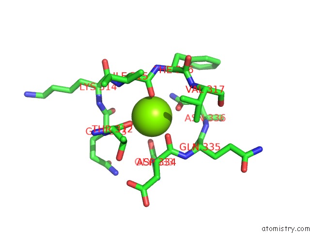
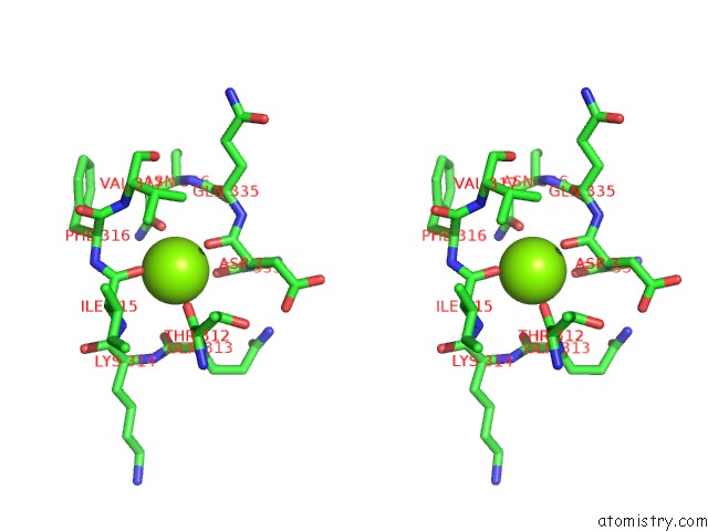
Magnesium binding site 1 out of 3 in 6rux
Go back to
Magnesium binding site 1 out
of 3 in the P46, An Immunodominant Surface Protein From Mycoplasma Hyopneumoniae
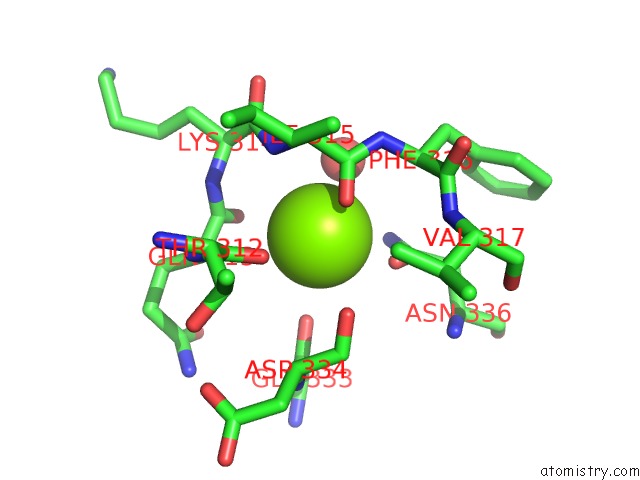
Mono view
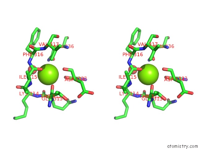
Stereo pair view
Mono view
Stereo pair view
A full contact list of Magnesium with other atoms in the Mg binding
site number 1 of P46, An Immunodominant Surface Protein From Mycoplasma Hyopneumoniae within 5.0Å range:
|
Magnesium binding site 2 out of 3 in 6rux
Go back to
Magnesium binding site 2 out
of 3 in the P46, An Immunodominant Surface Protein From Mycoplasma Hyopneumoniae
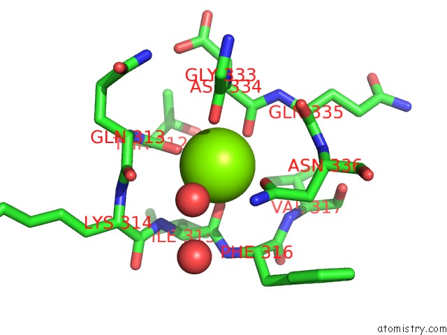
Mono view
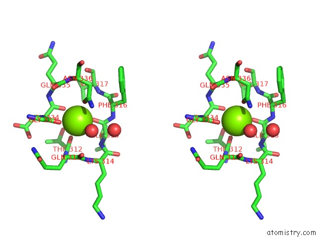
Stereo pair view
Mono view
Stereo pair view
A full contact list of Magnesium with other atoms in the Mg binding
site number 2 of P46, An Immunodominant Surface Protein From Mycoplasma Hyopneumoniae within 5.0Å range:
|
Magnesium binding site 3 out of 3 in 6rux
Go back to
Magnesium binding site 3 out
of 3 in the P46, An Immunodominant Surface Protein From Mycoplasma Hyopneumoniae
Mono view
Stereo pair view
Mono view
Stereo pair view
A full contact list of Magnesium with other atoms in the Mg binding
site number 3 of P46, An Immunodominant Surface Protein From Mycoplasma Hyopneumoniae within 5.0Å range:
|
Reference:
A.Guasch,
I.Fita.
Structure of P46 with Maltose To Be Published.
Page generated: Wed Aug 13 16:06:56 2025
Last articles
Mg in 7FS2Mg in 7FS1
Mg in 7FS0
Mg in 7FRZ
Mg in 7FRY
Mg in 7FRX
Mg in 7FRW
Mg in 7FRV
Mg in 7FJP
Mg in 7FQJ