Magnesium »
PDB 6s5d-6sh6 »
6s83 »
Magnesium in PDB 6s83: Crystal Structure of Methionine Adenosyltransferase From Pyrococcus Furiosus in Complex with Amppcp, Sam, and Pcp
Enzymatic activity of Crystal Structure of Methionine Adenosyltransferase From Pyrococcus Furiosus in Complex with Amppcp, Sam, and Pcp
All present enzymatic activity of Crystal Structure of Methionine Adenosyltransferase From Pyrococcus Furiosus in Complex with Amppcp, Sam, and Pcp:
2.5.1.6;
2.5.1.6;
Protein crystallography data
The structure of Crystal Structure of Methionine Adenosyltransferase From Pyrococcus Furiosus in Complex with Amppcp, Sam, and Pcp, PDB code: 6s83
was solved by
M.Degano,
C.Minici,
M.Porcelli,
with X-Ray Crystallography technique. A brief refinement statistics is given in the table below:
| Resolution Low / High (Å) | 74.47 / 2.34 |
| Space group | P 21 21 21 |
| Cell size a, b, c (Å), α, β, γ (°) | 78.196, 111.430, 400.400, 90.00, 90.00, 90.00 |
| R / Rfree (%) | 20.7 / 24 |
Magnesium Binding Sites:
Pages:
>>> Page 1 <<< Page 2, Binding sites: 11 - 20; Page 3, Binding sites: 21 - 21;Binding sites:
The binding sites of Magnesium atom in the Crystal Structure of Methionine Adenosyltransferase From Pyrococcus Furiosus in Complex with Amppcp, Sam, and Pcp (pdb code 6s83). This binding sites where shown within 5.0 Angstroms radius around Magnesium atom.In total 21 binding sites of Magnesium where determined in the Crystal Structure of Methionine Adenosyltransferase From Pyrococcus Furiosus in Complex with Amppcp, Sam, and Pcp, PDB code: 6s83:
Jump to Magnesium binding site number: 1; 2; 3; 4; 5; 6; 7; 8; 9; 10;
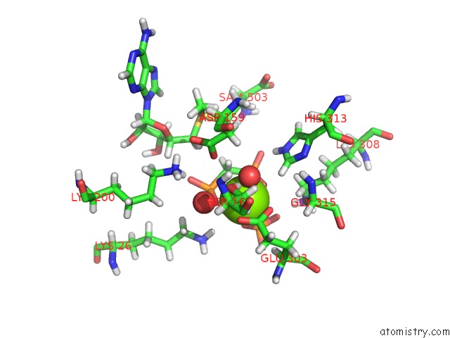
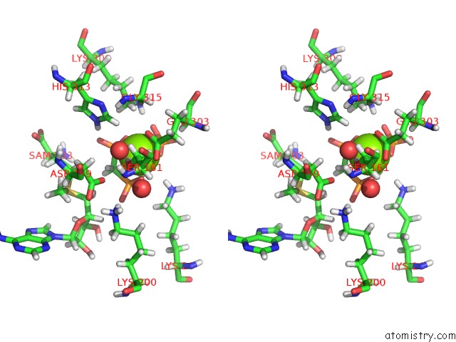
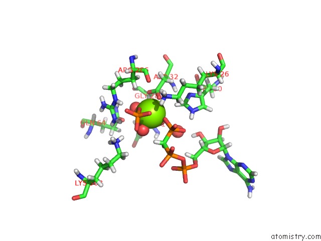
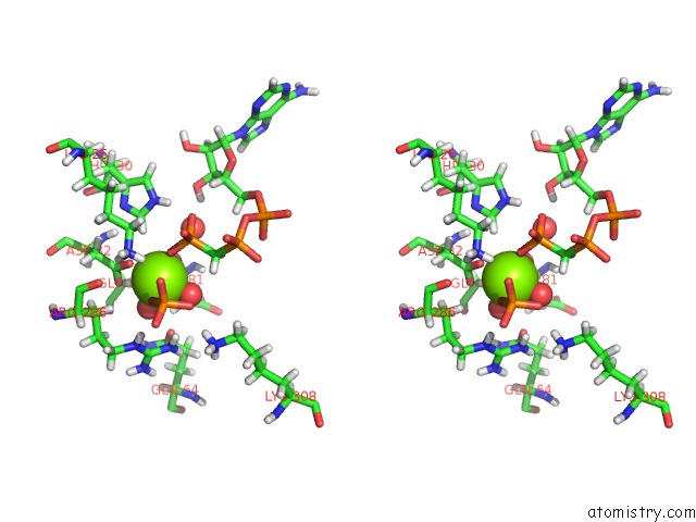
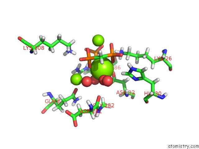
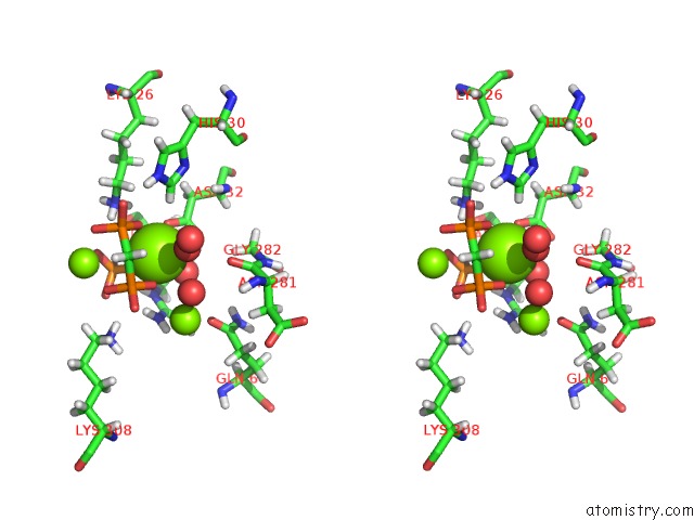
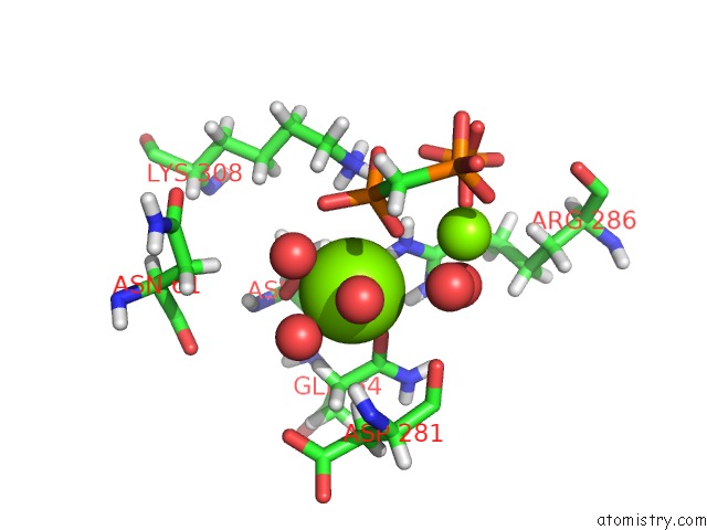
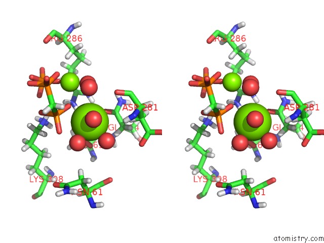
Magnesium binding site 1 out of 21 in 6s83
Go back to
Magnesium binding site 1 out
of 21 in the Crystal Structure of Methionine Adenosyltransferase From Pyrococcus Furiosus in Complex with Amppcp, Sam, and Pcp
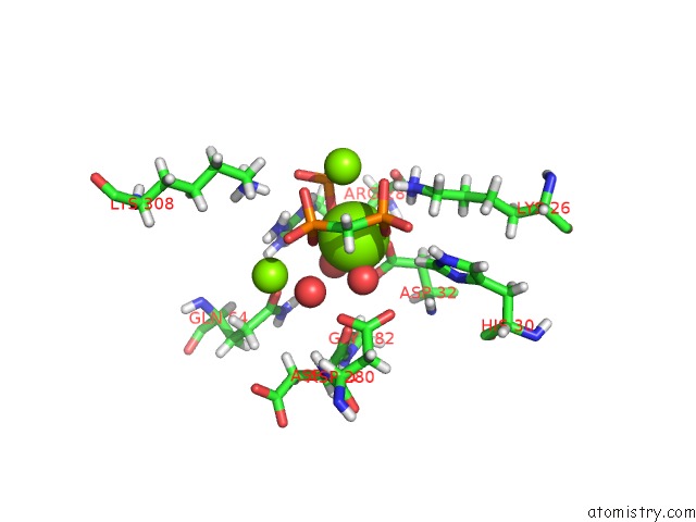
Mono view
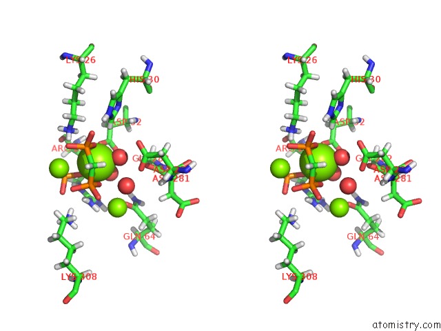
Stereo pair view
Mono view
Stereo pair view
A full contact list of Magnesium with other atoms in the Mg binding
site number 1 of Crystal Structure of Methionine Adenosyltransferase From Pyrococcus Furiosus in Complex with Amppcp, Sam, and Pcp within 5.0Å range:
|
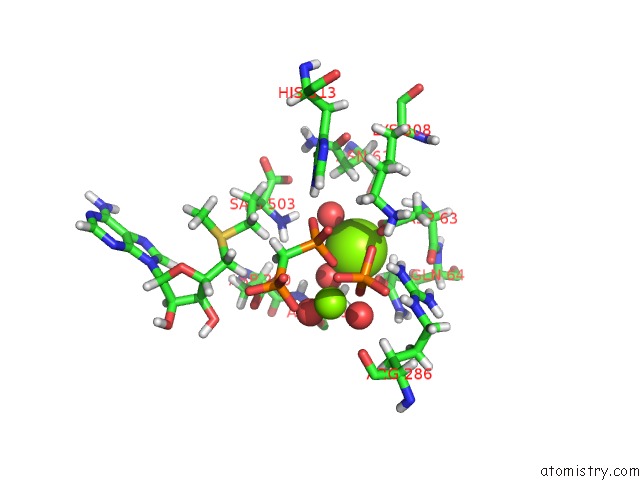
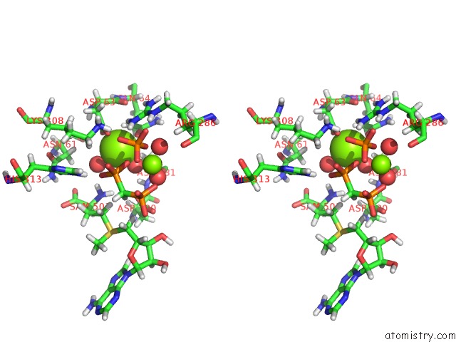
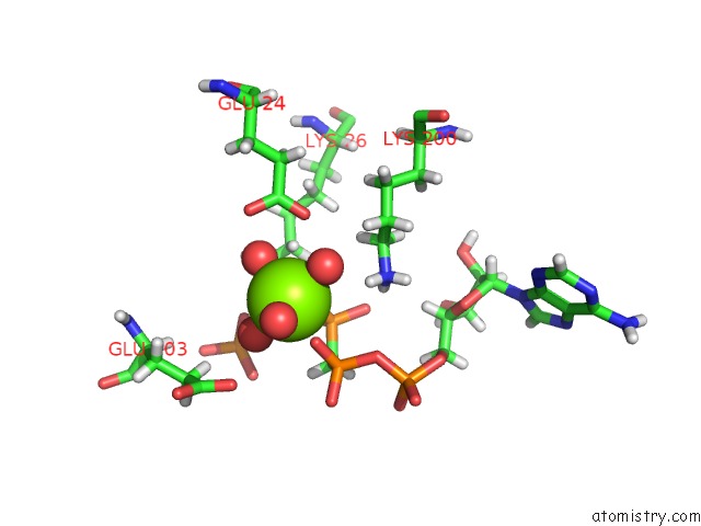
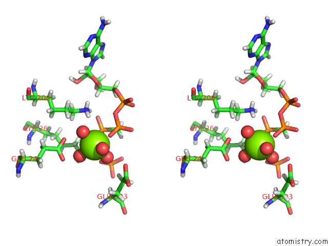
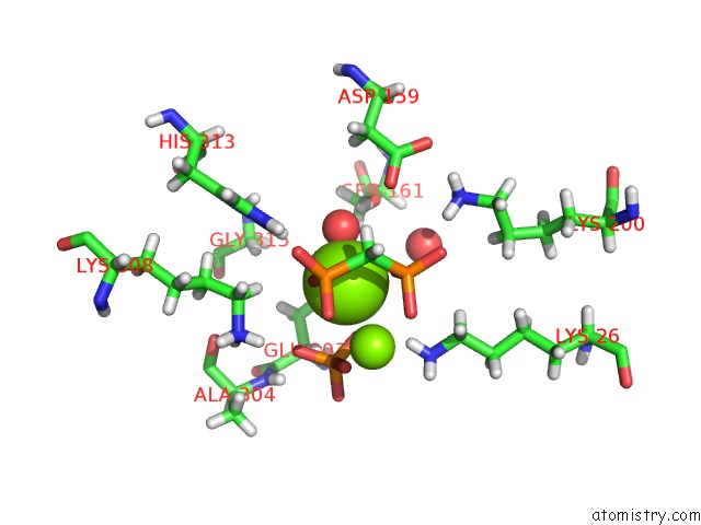
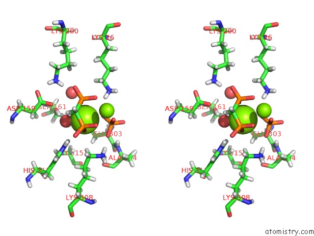
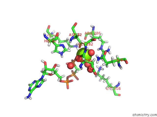
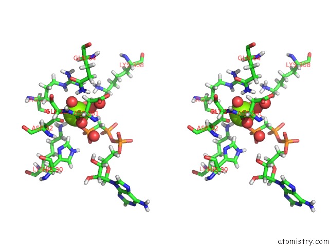
Magnesium binding site 2 out of 21 in 6s83
Go back to
Magnesium binding site 2 out
of 21 in the Crystal Structure of Methionine Adenosyltransferase From Pyrococcus Furiosus in Complex with Amppcp, Sam, and Pcp
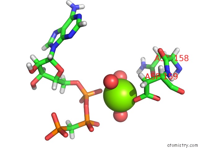
Mono view
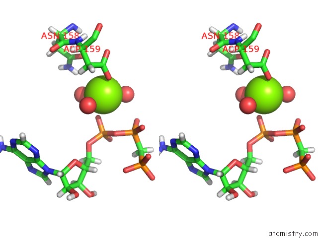
Stereo pair view
Mono view
Stereo pair view
A full contact list of Magnesium with other atoms in the Mg binding
site number 2 of Crystal Structure of Methionine Adenosyltransferase From Pyrococcus Furiosus in Complex with Amppcp, Sam, and Pcp within 5.0Å range:
|
Magnesium binding site 3 out of 21 in 6s83
Go back to
Magnesium binding site 3 out
of 21 in the Crystal Structure of Methionine Adenosyltransferase From Pyrococcus Furiosus in Complex with Amppcp, Sam, and Pcp
Mono view
Stereo pair view
Mono view
Stereo pair view
A full contact list of Magnesium with other atoms in the Mg binding
site number 3 of Crystal Structure of Methionine Adenosyltransferase From Pyrococcus Furiosus in Complex with Amppcp, Sam, and Pcp within 5.0Å range:
|
Magnesium binding site 4 out of 21 in 6s83
Go back to
Magnesium binding site 4 out
of 21 in the Crystal Structure of Methionine Adenosyltransferase From Pyrococcus Furiosus in Complex with Amppcp, Sam, and Pcp
Mono view
Stereo pair view
Mono view
Stereo pair view
A full contact list of Magnesium with other atoms in the Mg binding
site number 4 of Crystal Structure of Methionine Adenosyltransferase From Pyrococcus Furiosus in Complex with Amppcp, Sam, and Pcp within 5.0Å range:
|
Magnesium binding site 5 out of 21 in 6s83
Go back to
Magnesium binding site 5 out
of 21 in the Crystal Structure of Methionine Adenosyltransferase From Pyrococcus Furiosus in Complex with Amppcp, Sam, and Pcp
Mono view
Stereo pair view
Mono view
Stereo pair view
A full contact list of Magnesium with other atoms in the Mg binding
site number 5 of Crystal Structure of Methionine Adenosyltransferase From Pyrococcus Furiosus in Complex with Amppcp, Sam, and Pcp within 5.0Å range:
|
Magnesium binding site 6 out of 21 in 6s83
Go back to
Magnesium binding site 6 out
of 21 in the Crystal Structure of Methionine Adenosyltransferase From Pyrococcus Furiosus in Complex with Amppcp, Sam, and Pcp
Mono view
Stereo pair view
Mono view
Stereo pair view
A full contact list of Magnesium with other atoms in the Mg binding
site number 6 of Crystal Structure of Methionine Adenosyltransferase From Pyrococcus Furiosus in Complex with Amppcp, Sam, and Pcp within 5.0Å range:
|
Magnesium binding site 7 out of 21 in 6s83
Go back to
Magnesium binding site 7 out
of 21 in the Crystal Structure of Methionine Adenosyltransferase From Pyrococcus Furiosus in Complex with Amppcp, Sam, and Pcp
Mono view
Stereo pair view
Mono view
Stereo pair view
A full contact list of Magnesium with other atoms in the Mg binding
site number 7 of Crystal Structure of Methionine Adenosyltransferase From Pyrococcus Furiosus in Complex with Amppcp, Sam, and Pcp within 5.0Å range:
|
Magnesium binding site 8 out of 21 in 6s83
Go back to
Magnesium binding site 8 out
of 21 in the Crystal Structure of Methionine Adenosyltransferase From Pyrococcus Furiosus in Complex with Amppcp, Sam, and Pcp
Mono view
Stereo pair view
Mono view
Stereo pair view
A full contact list of Magnesium with other atoms in the Mg binding
site number 8 of Crystal Structure of Methionine Adenosyltransferase From Pyrococcus Furiosus in Complex with Amppcp, Sam, and Pcp within 5.0Å range:
|
Magnesium binding site 9 out of 21 in 6s83
Go back to
Magnesium binding site 9 out
of 21 in the Crystal Structure of Methionine Adenosyltransferase From Pyrococcus Furiosus in Complex with Amppcp, Sam, and Pcp
Mono view
Stereo pair view
Mono view
Stereo pair view
A full contact list of Magnesium with other atoms in the Mg binding
site number 9 of Crystal Structure of Methionine Adenosyltransferase From Pyrococcus Furiosus in Complex with Amppcp, Sam, and Pcp within 5.0Å range:
|
Magnesium binding site 10 out of 21 in 6s83
Go back to
Magnesium binding site 10 out
of 21 in the Crystal Structure of Methionine Adenosyltransferase From Pyrococcus Furiosus in Complex with Amppcp, Sam, and Pcp
Mono view
Stereo pair view
Mono view
Stereo pair view
A full contact list of Magnesium with other atoms in the Mg binding
site number 10 of Crystal Structure of Methionine Adenosyltransferase From Pyrococcus Furiosus in Complex with Amppcp, Sam, and Pcp within 5.0Å range:
|
Reference:
C.Minici,
L.Mosca,
C.Paola Ilisso,
G.Cacciapuoti,
M.Porcelli,
M.Degano.
Structures of Catalytic Cycle Intermediates of the Pyrococcus Furiosus Methionine Adenosyltransferase Demonstrate Negative Cooperativity in the Archaeal Orthologues. J.Struct.Biol. 07462 2020.
ISSN: ESSN 1095-8657
PubMed: 31962159
DOI: 10.1016/J.JSB.2020.107462
Page generated: Tue Oct 1 17:48:35 2024
ISSN: ESSN 1095-8657
PubMed: 31962159
DOI: 10.1016/J.JSB.2020.107462
Last articles
Zn in 9MJ5Zn in 9HNW
Zn in 9G0L
Zn in 9FNE
Zn in 9DZN
Zn in 9E0I
Zn in 9D32
Zn in 9DAK
Zn in 8ZXC
Zn in 8ZUF