Magnesium »
PDB 6sh6-6st5 »
6sjd »
Magnesium in PDB 6sjd: ZC3H12B-Ribonuclease Domain Bound to Rna
Protein crystallography data
The structure of ZC3H12B-Ribonuclease Domain Bound to Rna, PDB code: 6sjd
was solved by
E.Morgunova,
G.Bourenkov,
J.Taipale,
with X-Ray Crystallography technique. A brief refinement statistics is given in the table below:
| Resolution Low / High (Å) | 66.95 / 3.29 |
| Space group | P 43 21 2 |
| Cell size a, b, c (Å), α, β, γ (°) | 114.237, 114.237, 165.268, 90.00, 90.00, 90.00 |
| R / Rfree (%) | 23.5 / 27.9 |
Other elements in 6sjd:
The structure of ZC3H12B-Ribonuclease Domain Bound to Rna also contains other interesting chemical elements:
| Arsenic | (As) | 1 atom |
Magnesium Binding Sites:
The binding sites of Magnesium atom in the ZC3H12B-Ribonuclease Domain Bound to Rna
(pdb code 6sjd). This binding sites where shown within
5.0 Angstroms radius around Magnesium atom.
In total 2 binding sites of Magnesium where determined in the ZC3H12B-Ribonuclease Domain Bound to Rna, PDB code: 6sjd:
Jump to Magnesium binding site number: 1; 2;
In total 2 binding sites of Magnesium where determined in the ZC3H12B-Ribonuclease Domain Bound to Rna, PDB code: 6sjd:
Jump to Magnesium binding site number: 1; 2;
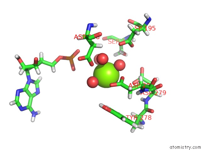
Magnesium binding site 1 out of 2 in 6sjd
Go back to
Magnesium binding site 1 out
of 2 in the ZC3H12B-Ribonuclease Domain Bound to Rna
Mono view

Stereo pair view
Mono view
Stereo pair view
A full contact list of Magnesium with other atoms in the Mg binding
site number 1 of ZC3H12B-Ribonuclease Domain Bound to Rna within 5.0Å range:
|
Magnesium binding site 2 out of 2 in 6sjd
Go back to
Magnesium binding site 2 out
of 2 in the ZC3H12B-Ribonuclease Domain Bound to Rna

Mono view

Stereo pair view
Mono view
Stereo pair view
A full contact list of Magnesium with other atoms in the Mg binding
site number 2 of ZC3H12B-Ribonuclease Domain Bound to Rna within 5.0Å range:
|
Reference:
A.Jolma,
J.Zhang,
E.Mondragon,
E.Morgunova,
T.Kivioja,
K.U.Laverty,
Y.Yin,
F.Zhu,
G.Bourenkov,
Q.Morris,
T.R.Hughes,
L.J.Maher 3Rd,
J.Taipale.
Binding Specificities of Human Rna-Binding Proteins Toward Structured and Linear Rna Sequences. Genome Res. V. 30 962 2020.
ISSN: ISSN 1088-9051
PubMed: 32703884
DOI: 10.1101/GR.258848.119
Page generated: Tue Oct 1 17:57:45 2024
ISSN: ISSN 1088-9051
PubMed: 32703884
DOI: 10.1101/GR.258848.119
Last articles
Zn in 9J0NZn in 9J0O
Zn in 9J0P
Zn in 9FJX
Zn in 9EKB
Zn in 9C0F
Zn in 9CAH
Zn in 9CH0
Zn in 9CH3
Zn in 9CH1