Magnesium »
PDB 6tfw-6tse »
6tqb »
Magnesium in PDB 6tqb: X-Ray Structure of Roquin Roq Domain in Complex with A UCP3 CDE1 Sl Rna Motif
Enzymatic activity of X-Ray Structure of Roquin Roq Domain in Complex with A UCP3 CDE1 Sl Rna Motif
All present enzymatic activity of X-Ray Structure of Roquin Roq Domain in Complex with A UCP3 CDE1 Sl Rna Motif:
2.3.2.27;
2.3.2.27;
Protein crystallography data
The structure of X-Ray Structure of Roquin Roq Domain in Complex with A UCP3 CDE1 Sl Rna Motif, PDB code: 6tqb
was solved by
O.Binas,
J.-N.Tants,
S.A.Peter,
R.Janowski,
E.Davydova,
J.Braun,
D.Niessing,
H.Schwalbe,
J.E.Weigand,
A.Schlundt,
with X-Ray Crystallography technique. A brief refinement statistics is given in the table below:
| Resolution Low / High (Å) | 47.08 / 1.60 |
| Space group | P 42 21 2 |
| Cell size a, b, c (Å), α, β, γ (°) | 86.990, 86.990, 72.990, 90.00, 90.00, 90.00 |
| R / Rfree (%) | 15.6 / 19.7 |
Other elements in 6tqb:
The structure of X-Ray Structure of Roquin Roq Domain in Complex with A UCP3 CDE1 Sl Rna Motif also contains other interesting chemical elements:
| Chlorine | (Cl) | 4 atoms |
| Sodium | (Na) | 2 atoms |
Magnesium Binding Sites:
The binding sites of Magnesium atom in the X-Ray Structure of Roquin Roq Domain in Complex with A UCP3 CDE1 Sl Rna Motif
(pdb code 6tqb). This binding sites where shown within
5.0 Angstroms radius around Magnesium atom.
In total only one binding site of Magnesium was determined in the X-Ray Structure of Roquin Roq Domain in Complex with A UCP3 CDE1 Sl Rna Motif, PDB code: 6tqb:
In total only one binding site of Magnesium was determined in the X-Ray Structure of Roquin Roq Domain in Complex with A UCP3 CDE1 Sl Rna Motif, PDB code: 6tqb:
Magnesium binding site 1 out of 1 in 6tqb
Go back to
Magnesium binding site 1 out
of 1 in the X-Ray Structure of Roquin Roq Domain in Complex with A UCP3 CDE1 Sl Rna Motif
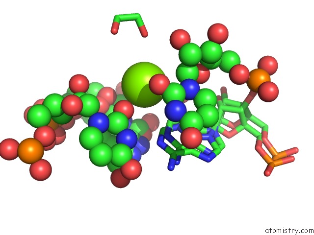
Mono view
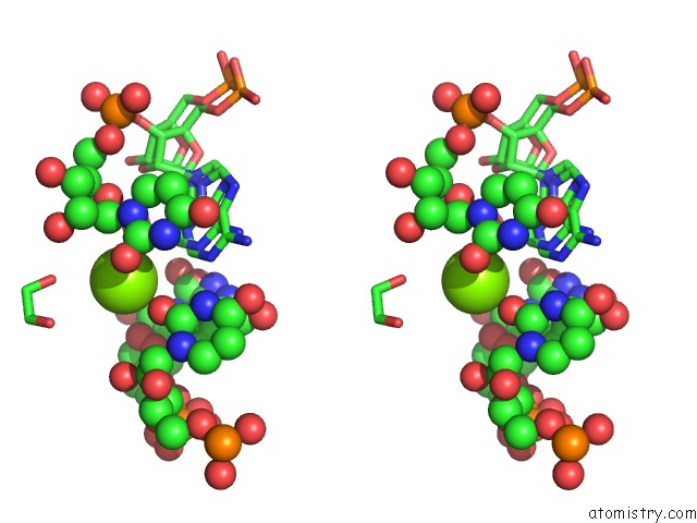
Stereo pair view
Mono view
Stereo pair view
A full contact list of Magnesium with other atoms in the Mg binding
site number 1 of X-Ray Structure of Roquin Roq Domain in Complex with A UCP3 CDE1 Sl Rna Motif within 5.0Å range:
|
Reference:
R.Janowski,
E.Davydova,
A.Schlundt,
D.Niessing.
Structural Basis For the Recognition of (Linear and) Non-Linear Au-Rich Elements By Roquin To Be Published.
Page generated: Tue Oct 1 19:04:37 2024
Last articles
Fe in 2YXOFe in 2YRS
Fe in 2YXC
Fe in 2YNM
Fe in 2YVJ
Fe in 2YP1
Fe in 2YU2
Fe in 2YU1
Fe in 2YQB
Fe in 2YOO