Magnesium »
PDB 6u1y-6ufh »
6u3j »
Magnesium in PDB 6u3j: Structure of the 2-Oxoadipate Dehydrogenase DHTKD1
Enzymatic activity of Structure of the 2-Oxoadipate Dehydrogenase DHTKD1
All present enzymatic activity of Structure of the 2-Oxoadipate Dehydrogenase DHTKD1:
1.2.4.2;
1.2.4.2;
Protein crystallography data
The structure of Structure of the 2-Oxoadipate Dehydrogenase DHTKD1, PDB code: 6u3j
was solved by
S.Khamrui,
M.B.Lazarus,
with X-Ray Crystallography technique. A brief refinement statistics is given in the table below:
| Resolution Low / High (Å) | 49.01 / 2.25 |
| Space group | C 1 2 1 |
| Cell size a, b, c (Å), α, β, γ (°) | 332.314, 72.612, 79.675, 90.00, 91.87, 90.00 |
| R / Rfree (%) | 20.9 / 24.1 |
Magnesium Binding Sites:
The binding sites of Magnesium atom in the Structure of the 2-Oxoadipate Dehydrogenase DHTKD1
(pdb code 6u3j). This binding sites where shown within
5.0 Angstroms radius around Magnesium atom.
In total 3 binding sites of Magnesium where determined in the Structure of the 2-Oxoadipate Dehydrogenase DHTKD1, PDB code: 6u3j:
Jump to Magnesium binding site number: 1; 2; 3;
In total 3 binding sites of Magnesium where determined in the Structure of the 2-Oxoadipate Dehydrogenase DHTKD1, PDB code: 6u3j:
Jump to Magnesium binding site number: 1; 2; 3;
Magnesium binding site 1 out of 3 in 6u3j
Go back to
Magnesium binding site 1 out
of 3 in the Structure of the 2-Oxoadipate Dehydrogenase DHTKD1
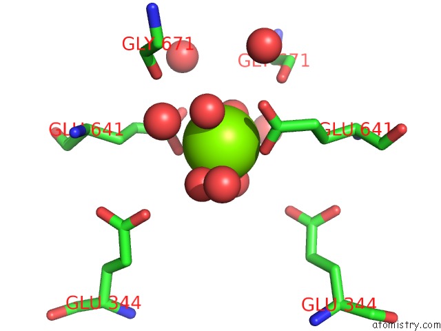
Mono view
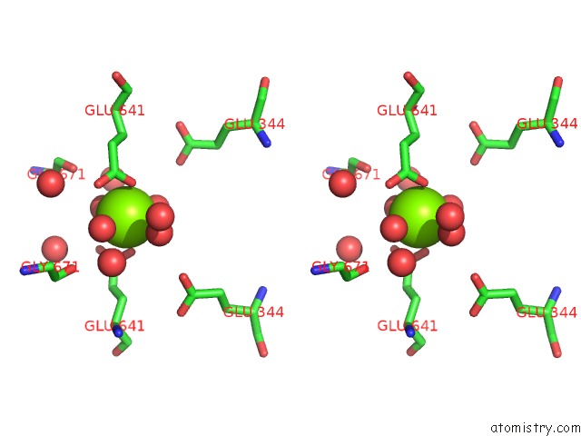
Stereo pair view
Mono view
Stereo pair view
A full contact list of Magnesium with other atoms in the Mg binding
site number 1 of Structure of the 2-Oxoadipate Dehydrogenase DHTKD1 within 5.0Å range:
|
Magnesium binding site 2 out of 3 in 6u3j
Go back to
Magnesium binding site 2 out
of 3 in the Structure of the 2-Oxoadipate Dehydrogenase DHTKD1

Mono view

Stereo pair view
Mono view
Stereo pair view
A full contact list of Magnesium with other atoms in the Mg binding
site number 2 of Structure of the 2-Oxoadipate Dehydrogenase DHTKD1 within 5.0Å range:
|
Magnesium binding site 3 out of 3 in 6u3j
Go back to
Magnesium binding site 3 out
of 3 in the Structure of the 2-Oxoadipate Dehydrogenase DHTKD1

Mono view

Stereo pair view
Mono view
Stereo pair view
A full contact list of Magnesium with other atoms in the Mg binding
site number 3 of Structure of the 2-Oxoadipate Dehydrogenase DHTKD1 within 5.0Å range:
|
Reference:
J.Leandro,
S.Khamrui,
H.Wang,
C.Suebsuwong,
N.S.Nemeria,
K.Huynh,
M.Moustakim,
C.Secor,
M.Wang,
T.Dodatko,
B.Stauffer,
C.G.Wilson,
C.Yu,
M.R.Arkin,
F.Jordan,
R.Sanchez,
R.J.Devita,
M.B.Lazarus,
S.M.Houten.
Inhibition and Crystal Structure of the Human DHTKD1-Thiamin Diphosphate Complex. Acs Chem.Biol. V. 15 2041 2020.
ISSN: ESSN 1554-8937
PubMed: 32633484
DOI: 10.1021/ACSCHEMBIO.0C00114
Page generated: Tue Oct 1 20:49:08 2024
ISSN: ESSN 1554-8937
PubMed: 32633484
DOI: 10.1021/ACSCHEMBIO.0C00114
Last articles
Zn in 9MJ5Zn in 9HNW
Zn in 9G0L
Zn in 9FNE
Zn in 9DZN
Zn in 9E0I
Zn in 9D32
Zn in 9DAK
Zn in 8ZXC
Zn in 8ZUF