Magnesium »
PDB 6u1y-6ufh »
6uec »
Magnesium in PDB 6uec: Pseudomonas Aeruginosa Lpxd Complex Structure with Ligand
Protein crystallography data
The structure of Pseudomonas Aeruginosa Lpxd Complex Structure with Ligand, PDB code: 6uec
was solved by
Y.Chen,
K.Kroeck,
M.Sacco,
with X-Ray Crystallography technique. A brief refinement statistics is given in the table below:
| Resolution Low / High (Å) | 32.99 / 2.60 |
| Space group | H 3 |
| Cell size a, b, c (Å), α, β, γ (°) | 104.764, 104.764, 95.967, 90.00, 90.00, 120.00 |
| R / Rfree (%) | 20.9 / 28.9 |
Magnesium Binding Sites:
The binding sites of Magnesium atom in the Pseudomonas Aeruginosa Lpxd Complex Structure with Ligand
(pdb code 6uec). This binding sites where shown within
5.0 Angstroms radius around Magnesium atom.
In total only one binding site of Magnesium was determined in the Pseudomonas Aeruginosa Lpxd Complex Structure with Ligand, PDB code: 6uec:
In total only one binding site of Magnesium was determined in the Pseudomonas Aeruginosa Lpxd Complex Structure with Ligand, PDB code: 6uec:
Magnesium binding site 1 out of 1 in 6uec
Go back to
Magnesium binding site 1 out
of 1 in the Pseudomonas Aeruginosa Lpxd Complex Structure with Ligand
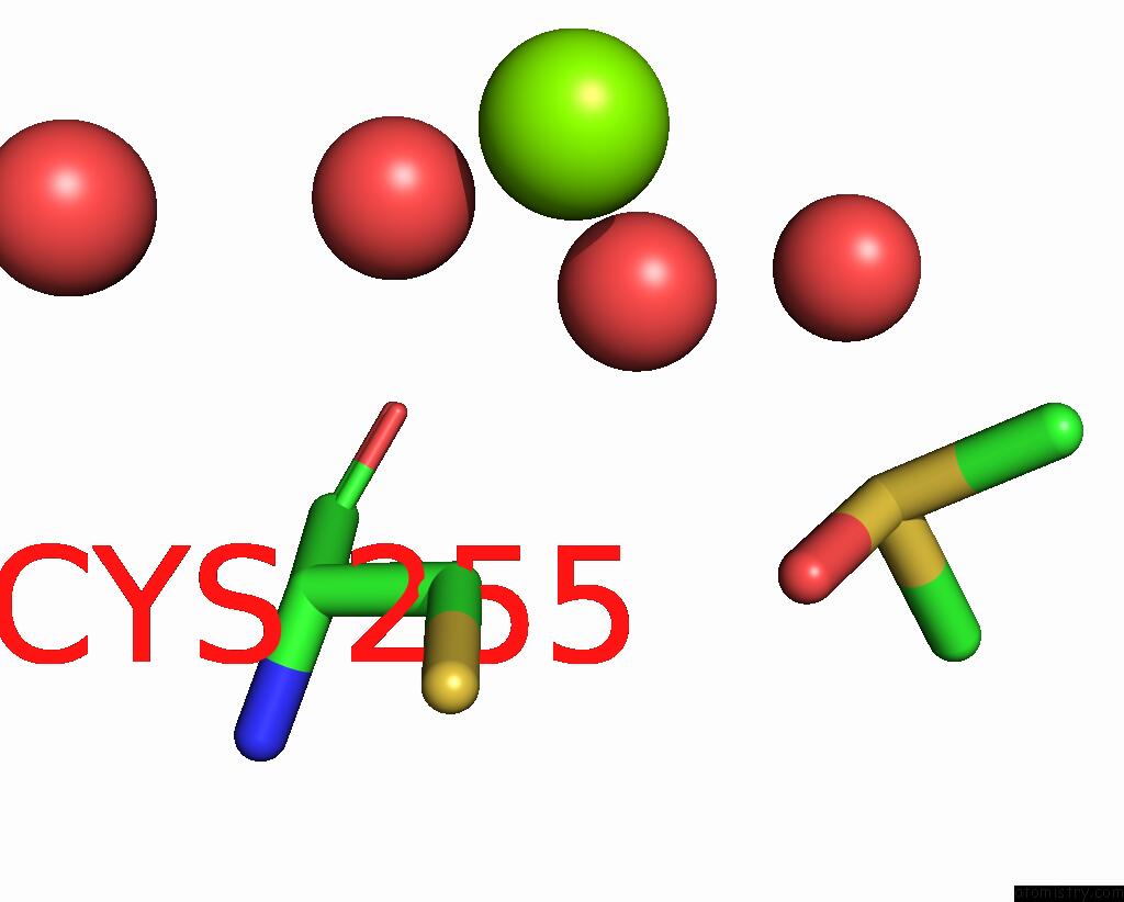
Mono view
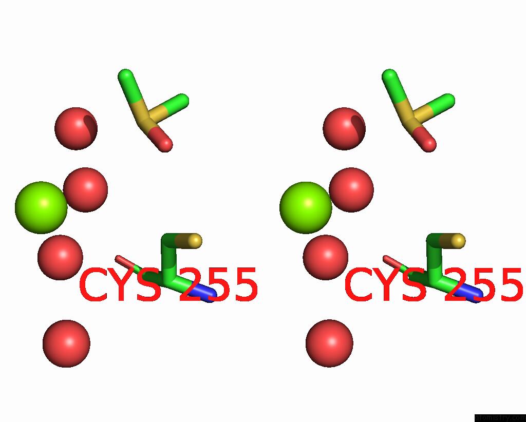
Stereo pair view
Mono view
Stereo pair view
A full contact list of Magnesium with other atoms in the Mg binding
site number 1 of Pseudomonas Aeruginosa Lpxd Complex Structure with Ligand within 5.0Å range:
|
Reference:
K.G.Kroeck,
M.D.Sacco,
E.W.Smith,
X.Zhang,
D.Shoun,
A.Akhtar,
S.E.Darch,
F.Cohen,
L.D.Andrews,
J.E.Knox,
Y.Chen.
Discovery of Dual-Activity Small-Molecule Ligands of Pseudomonas Aeruginosa Lpxa and Lpxd Using Spr and X-Ray Crystallography. Sci Rep V. 9 15450 2019.
ISSN: ESSN 2045-2322
PubMed: 31664082
DOI: 10.1038/S41598-019-51844-Z
Page generated: Tue Oct 1 20:58:52 2024
ISSN: ESSN 2045-2322
PubMed: 31664082
DOI: 10.1038/S41598-019-51844-Z
Last articles
Ca in 5SZNCa in 5SZM
Ca in 5SZL
Ca in 5SY1
Ca in 5SWI
Ca in 5SVE
Ca in 5SSX
Ca in 5SV0
Ca in 5STD
Ca in 5SSZ