Magnesium »
PDB 6ufg-6uok »
6ui2 »
Magnesium in PDB 6ui2: Structure of Human Dna Polymerase Eta Complexed with N7MG in the Template Base Paired with Incoming Non-Hydrolyzable Ctp
Enzymatic activity of Structure of Human Dna Polymerase Eta Complexed with N7MG in the Template Base Paired with Incoming Non-Hydrolyzable Ctp
All present enzymatic activity of Structure of Human Dna Polymerase Eta Complexed with N7MG in the Template Base Paired with Incoming Non-Hydrolyzable Ctp:
2.7.7.7;
2.7.7.7;
Protein crystallography data
The structure of Structure of Human Dna Polymerase Eta Complexed with N7MG in the Template Base Paired with Incoming Non-Hydrolyzable Ctp, PDB code: 6ui2
was solved by
M.C.Koag,
S.Lee,
with X-Ray Crystallography technique. A brief refinement statistics is given in the table below:
| Resolution Low / High (Å) | 19.90 / 2.35 |
| Space group | P 61 |
| Cell size a, b, c (Å), α, β, γ (°) | 98.684, 98.684, 81.852, 90.00, 90.00, 120.00 |
| R / Rfree (%) | 17 / 23.7 |
Other elements in 6ui2:
The structure of Structure of Human Dna Polymerase Eta Complexed with N7MG in the Template Base Paired with Incoming Non-Hydrolyzable Ctp also contains other interesting chemical elements:
| Fluorine | (F) | 1 atom |
Magnesium Binding Sites:
The binding sites of Magnesium atom in the Structure of Human Dna Polymerase Eta Complexed with N7MG in the Template Base Paired with Incoming Non-Hydrolyzable Ctp
(pdb code 6ui2). This binding sites where shown within
5.0 Angstroms radius around Magnesium atom.
In total 2 binding sites of Magnesium where determined in the Structure of Human Dna Polymerase Eta Complexed with N7MG in the Template Base Paired with Incoming Non-Hydrolyzable Ctp, PDB code: 6ui2:
Jump to Magnesium binding site number: 1; 2;
In total 2 binding sites of Magnesium where determined in the Structure of Human Dna Polymerase Eta Complexed with N7MG in the Template Base Paired with Incoming Non-Hydrolyzable Ctp, PDB code: 6ui2:
Jump to Magnesium binding site number: 1; 2;
Magnesium binding site 1 out of 2 in 6ui2
Go back to
Magnesium binding site 1 out
of 2 in the Structure of Human Dna Polymerase Eta Complexed with N7MG in the Template Base Paired with Incoming Non-Hydrolyzable Ctp
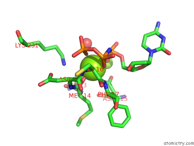
Mono view
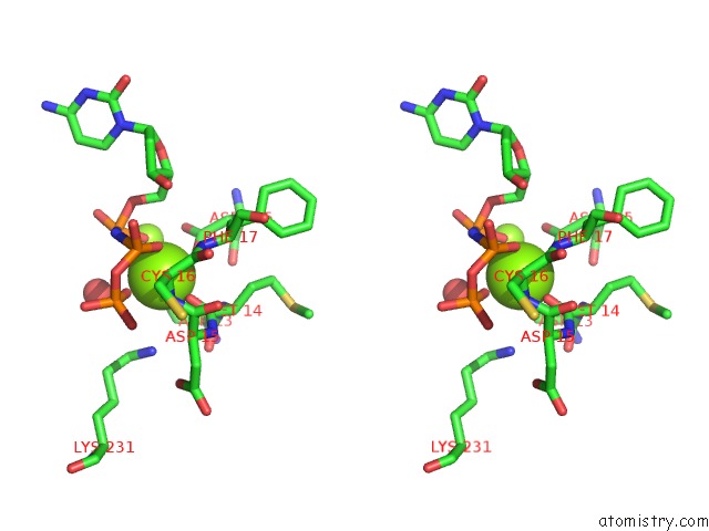
Stereo pair view
Mono view
Stereo pair view
A full contact list of Magnesium with other atoms in the Mg binding
site number 1 of Structure of Human Dna Polymerase Eta Complexed with N7MG in the Template Base Paired with Incoming Non-Hydrolyzable Ctp within 5.0Å range:
|
Magnesium binding site 2 out of 2 in 6ui2
Go back to
Magnesium binding site 2 out
of 2 in the Structure of Human Dna Polymerase Eta Complexed with N7MG in the Template Base Paired with Incoming Non-Hydrolyzable Ctp
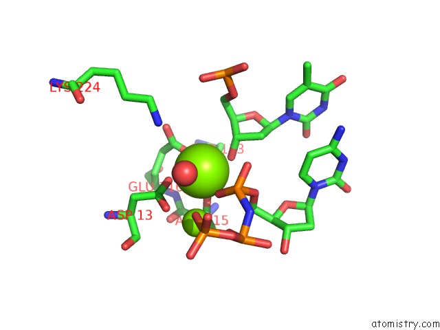
Mono view
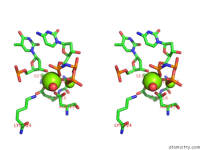
Stereo pair view
Mono view
Stereo pair view
A full contact list of Magnesium with other atoms in the Mg binding
site number 2 of Structure of Human Dna Polymerase Eta Complexed with N7MG in the Template Base Paired with Incoming Non-Hydrolyzable Ctp within 5.0Å range:
|
Reference:
M.C.Koag,
H.Jung,
Y.Kou,
S.Lee.
Bypass of the Major Alkylative Dna Lesion By Human Dna Polymerase Eta. Molecules V. 24 2019.
ISSN: ESSN 1420-3049
PubMed: 31683505
DOI: 10.3390/MOLECULES24213928
Page generated: Tue Oct 1 21:03:40 2024
ISSN: ESSN 1420-3049
PubMed: 31683505
DOI: 10.3390/MOLECULES24213928
Last articles
Zn in 9JYWZn in 9IR4
Zn in 9IR3
Zn in 9GMX
Zn in 9GMW
Zn in 9JEJ
Zn in 9ERF
Zn in 9ERE
Zn in 9EGV
Zn in 9EGW