Magnesium »
PDB 6ufh-6up0 »
6ui4 »
Magnesium in PDB 6ui4: Crystal Structure of Phenamacril-Bound F. Graminearum Myosin I
Protein crystallography data
The structure of Crystal Structure of Phenamacril-Bound F. Graminearum Myosin I, PDB code: 6ui4
was solved by
Y.Zhou,
X.E.Zhou,
Y.Gong,
Y.Zhu,
H.E.Xu,
M.Zhou,
K.Melcher,
F.Zhang,
with X-Ray Crystallography technique. A brief refinement statistics is given in the table below:
| Resolution Low / High (Å) | 42.91 / 2.65 |
| Space group | P 21 21 21 |
| Cell size a, b, c (Å), α, β, γ (°) | 59.502, 100.311, 165.615, 90.00, 90.00, 90.00 |
| R / Rfree (%) | 21.1 / 24.4 |
Magnesium Binding Sites:
The binding sites of Magnesium atom in the Crystal Structure of Phenamacril-Bound F. Graminearum Myosin I
(pdb code 6ui4). This binding sites where shown within
5.0 Angstroms radius around Magnesium atom.
In total only one binding site of Magnesium was determined in the Crystal Structure of Phenamacril-Bound F. Graminearum Myosin I, PDB code: 6ui4:
In total only one binding site of Magnesium was determined in the Crystal Structure of Phenamacril-Bound F. Graminearum Myosin I, PDB code: 6ui4:
Magnesium binding site 1 out of 1 in 6ui4
Go back to
Magnesium binding site 1 out
of 1 in the Crystal Structure of Phenamacril-Bound F. Graminearum Myosin I
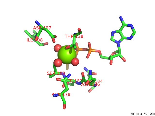
Mono view
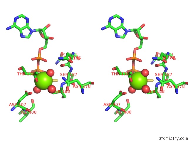
Stereo pair view
Mono view
Stereo pair view
A full contact list of Magnesium with other atoms in the Mg binding
site number 1 of Crystal Structure of Phenamacril-Bound F. Graminearum Myosin I within 5.0Å range:
|
Reference:
Y.Zhou,
X.E.Zhou,
Y.Gong,
Y.Zhu,
X.Cao,
J.S.Brunzelle,
H.E.Xu,
M.Zhou,
K.Melcher,
F.Zhang.
Structural Basis of Fusarium Myosin I Inhibition By Phenamacril. Plos Pathog. V. 16 08323 2020.
ISSN: ESSN 1553-7374
PubMed: 32163521
DOI: 10.1371/JOURNAL.PPAT.1008323
Page generated: Tue Oct 1 21:04:06 2024
ISSN: ESSN 1553-7374
PubMed: 32163521
DOI: 10.1371/JOURNAL.PPAT.1008323
Last articles
Zn in 9J0NZn in 9J0O
Zn in 9J0P
Zn in 9FJX
Zn in 9EKB
Zn in 9C0F
Zn in 9CAH
Zn in 9CH0
Zn in 9CH3
Zn in 9CH1