Magnesium »
PDB 6ufj-6up4 »
6uo6 »
Magnesium in PDB 6uo6: Crystal Structure of the R422Q Missense Variant of Human PGM1
Enzymatic activity of Crystal Structure of the R422Q Missense Variant of Human PGM1
All present enzymatic activity of Crystal Structure of the R422Q Missense Variant of Human PGM1:
5.4.2.2;
5.4.2.2;
Protein crystallography data
The structure of Crystal Structure of the R422Q Missense Variant of Human PGM1, PDB code: 6uo6
was solved by
L.J.Beamer,
K.M.Stiers,
with X-Ray Crystallography technique. A brief refinement statistics is given in the table below:
| Resolution Low / High (Å) | 49.61 / 2.15 |
| Space group | P 41 21 2 |
| Cell size a, b, c (Å), α, β, γ (°) | 171.804, 171.804, 99.278, 90.00, 90.00, 90.00 |
| R / Rfree (%) | 17.5 / 21.3 |
Magnesium Binding Sites:
The binding sites of Magnesium atom in the Crystal Structure of the R422Q Missense Variant of Human PGM1
(pdb code 6uo6). This binding sites where shown within
5.0 Angstroms radius around Magnesium atom.
In total 2 binding sites of Magnesium where determined in the Crystal Structure of the R422Q Missense Variant of Human PGM1, PDB code: 6uo6:
Jump to Magnesium binding site number: 1; 2;
In total 2 binding sites of Magnesium where determined in the Crystal Structure of the R422Q Missense Variant of Human PGM1, PDB code: 6uo6:
Jump to Magnesium binding site number: 1; 2;
Magnesium binding site 1 out of 2 in 6uo6
Go back to
Magnesium binding site 1 out
of 2 in the Crystal Structure of the R422Q Missense Variant of Human PGM1
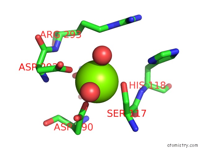
Mono view
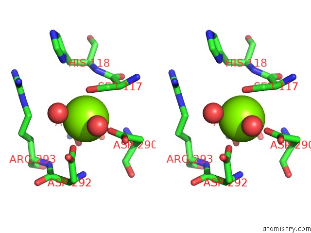
Stereo pair view
Mono view
Stereo pair view
A full contact list of Magnesium with other atoms in the Mg binding
site number 1 of Crystal Structure of the R422Q Missense Variant of Human PGM1 within 5.0Å range:
|
Magnesium binding site 2 out of 2 in 6uo6
Go back to
Magnesium binding site 2 out
of 2 in the Crystal Structure of the R422Q Missense Variant of Human PGM1
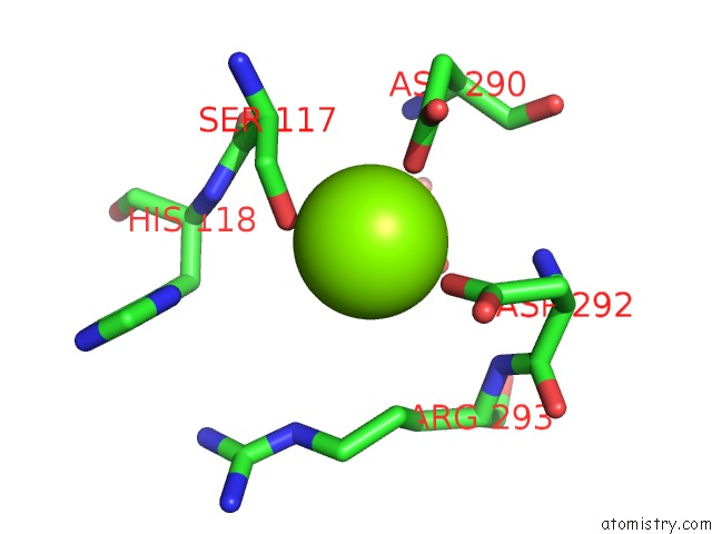
Mono view
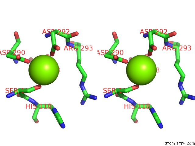
Stereo pair view
Mono view
Stereo pair view
A full contact list of Magnesium with other atoms in the Mg binding
site number 2 of Crystal Structure of the R422Q Missense Variant of Human PGM1 within 5.0Å range:
|
Reference:
K.M.Stiers,
R.P.Hansen,
B.A.Daghlas,
K.N.Mason,
J.S.Zhu,
D.L.Jakeman,
L.J.Beamer.
A Missense Variant Remote From the Active Site Impairs Stability of Human Phosphoglucomutase 1. J. Inherit. Metab. Dis. 2020.
ISSN: ISSN 1573-2665
PubMed: 32057119
DOI: 10.1002/JIMD.12222
Page generated: Tue Oct 1 21:08:51 2024
ISSN: ISSN 1573-2665
PubMed: 32057119
DOI: 10.1002/JIMD.12222
Last articles
Zn in 9MJ5Zn in 9HNW
Zn in 9G0L
Zn in 9FNE
Zn in 9DZN
Zn in 9E0I
Zn in 9D32
Zn in 9DAK
Zn in 8ZXC
Zn in 8ZUF