Magnesium »
PDB 6vpb-6vzt »
6vpm »
Magnesium in PDB 6vpm: TPX2 Residues 7-20 Fused to Aurora A Residues 116-389 with C290 Disulfide Bonded to Compound 8-34, and in Complex with Amp-Pnp
Enzymatic activity of TPX2 Residues 7-20 Fused to Aurora A Residues 116-389 with C290 Disulfide Bonded to Compound 8-34, and in Complex with Amp-Pnp
All present enzymatic activity of TPX2 Residues 7-20 Fused to Aurora A Residues 116-389 with C290 Disulfide Bonded to Compound 8-34, and in Complex with Amp-Pnp:
2.7.11.1;
2.7.11.1;
Protein crystallography data
The structure of TPX2 Residues 7-20 Fused to Aurora A Residues 116-389 with C290 Disulfide Bonded to Compound 8-34, and in Complex with Amp-Pnp, PDB code: 6vpm
was solved by
D.C.Lim,
M.B.Yaffe,
with X-Ray Crystallography technique. A brief refinement statistics is given in the table below:
| Resolution Low / High (Å) | 22.87 / 1.58 |
| Space group | P 21 21 21 |
| Cell size a, b, c (Å), α, β, γ (°) | 50.730, 85.769, 152.492, 90.00, 90.00, 90.00 |
| R / Rfree (%) | 17 / 20.3 |
Magnesium Binding Sites:
The binding sites of Magnesium atom in the TPX2 Residues 7-20 Fused to Aurora A Residues 116-389 with C290 Disulfide Bonded to Compound 8-34, and in Complex with Amp-Pnp
(pdb code 6vpm). This binding sites where shown within
5.0 Angstroms radius around Magnesium atom.
In total 2 binding sites of Magnesium where determined in the TPX2 Residues 7-20 Fused to Aurora A Residues 116-389 with C290 Disulfide Bonded to Compound 8-34, and in Complex with Amp-Pnp, PDB code: 6vpm:
Jump to Magnesium binding site number: 1; 2;
In total 2 binding sites of Magnesium where determined in the TPX2 Residues 7-20 Fused to Aurora A Residues 116-389 with C290 Disulfide Bonded to Compound 8-34, and in Complex with Amp-Pnp, PDB code: 6vpm:
Jump to Magnesium binding site number: 1; 2;
Magnesium binding site 1 out of 2 in 6vpm
Go back to
Magnesium binding site 1 out
of 2 in the TPX2 Residues 7-20 Fused to Aurora A Residues 116-389 with C290 Disulfide Bonded to Compound 8-34, and in Complex with Amp-Pnp
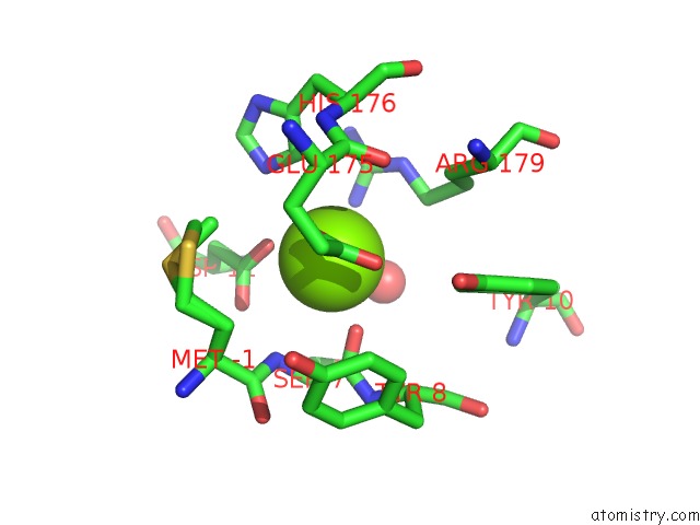
Mono view
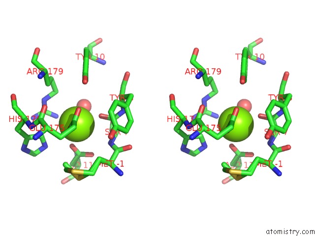
Stereo pair view
Mono view
Stereo pair view
A full contact list of Magnesium with other atoms in the Mg binding
site number 1 of TPX2 Residues 7-20 Fused to Aurora A Residues 116-389 with C290 Disulfide Bonded to Compound 8-34, and in Complex with Amp-Pnp within 5.0Å range:
|
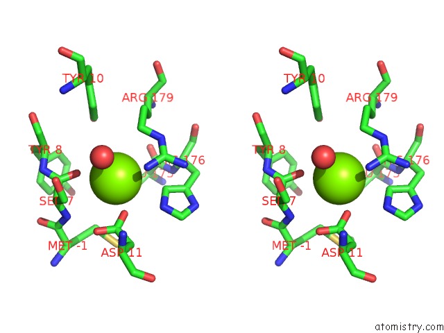
Magnesium binding site 2 out of 2 in 6vpm
Go back to
Magnesium binding site 2 out
of 2 in the TPX2 Residues 7-20 Fused to Aurora A Residues 116-389 with C290 Disulfide Bonded to Compound 8-34, and in Complex with Amp-Pnp

Mono view
Stereo pair view
Mono view
Stereo pair view
A full contact list of Magnesium with other atoms in the Mg binding
site number 2 of TPX2 Residues 7-20 Fused to Aurora A Residues 116-389 with C290 Disulfide Bonded to Compound 8-34, and in Complex with Amp-Pnp within 5.0Å range:
|
Reference:
D.C.Lim,
V.Joukov,
T.J.Rettenmaier,
A.Kumagai,
W.G.Dunphy,
J.A.Wells,
M.B.Yaffe.
Redox Priming Promotes Aurora A Activation During Mitosis. Sci.Signal. V. 13 2020.
ISSN: ESSN 1937-9145
PubMed: 32694171
DOI: 10.1126/SCISIGNAL.ABB6707
Page generated: Wed Aug 13 19:17:32 2025
ISSN: ESSN 1937-9145
PubMed: 32694171
DOI: 10.1126/SCISIGNAL.ABB6707
Last articles
Mg in 7AA2Mg in 7A8Q
Mg in 7A8R
Mg in 7A7E
Mg in 7A6H
Mg in 7A5Z
Mg in 7A5E
Mg in 7A2F
Mg in 7A2B
Mg in 7A3Z