Magnesium »
PDB 6voy-6vzr »
6vww »
Magnesium in PDB 6vww: Crystal Structure of NSP15 Endoribonuclease From Sars Cov-2.
Protein crystallography data
The structure of Crystal Structure of NSP15 Endoribonuclease From Sars Cov-2., PDB code: 6vww
was solved by
Y.Kim,
R.Jedrzejczak,
N.Maltseva,
M.Endres,
A.Godzik,
K.Michalska,
A.Joachimiak,
Center For Structural Genomics Of Infectious Diseases(Csgid),
with X-Ray Crystallography technique. A brief refinement statistics is given in the table below:
| Resolution Low / High (Å) | 45.06 / 2.20 |
| Space group | P 63 |
| Cell size a, b, c (Å), α, β, γ (°) | 150.539, 150.539, 111.310, 90.00, 90.00, 120.00 |
| R / Rfree (%) | 15.8 / 17.8 |
Other elements in 6vww:
The structure of Crystal Structure of NSP15 Endoribonuclease From Sars Cov-2. also contains other interesting chemical elements:
| Chlorine | (Cl) | 1 atom |
Magnesium Binding Sites:
The binding sites of Magnesium atom in the Crystal Structure of NSP15 Endoribonuclease From Sars Cov-2.
(pdb code 6vww). This binding sites where shown within
5.0 Angstroms radius around Magnesium atom.
In total only one binding site of Magnesium was determined in the Crystal Structure of NSP15 Endoribonuclease From Sars Cov-2., PDB code: 6vww:
In total only one binding site of Magnesium was determined in the Crystal Structure of NSP15 Endoribonuclease From Sars Cov-2., PDB code: 6vww:
Magnesium binding site 1 out of 1 in 6vww
Go back to
Magnesium binding site 1 out
of 1 in the Crystal Structure of NSP15 Endoribonuclease From Sars Cov-2.
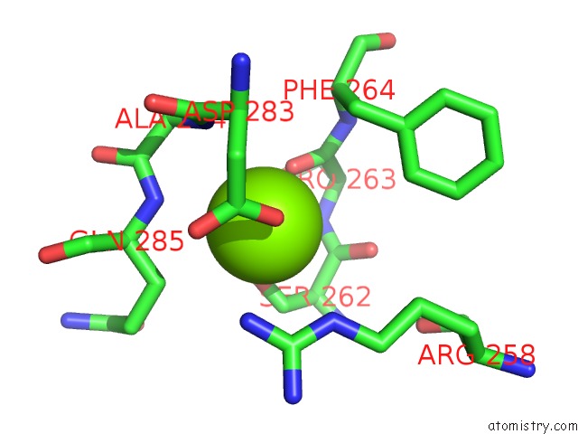
Mono view
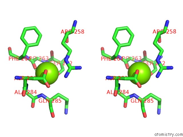
Stereo pair view
Mono view
Stereo pair view
A full contact list of Magnesium with other atoms in the Mg binding
site number 1 of Crystal Structure of NSP15 Endoribonuclease From Sars Cov-2. within 5.0Å range:
|
Reference:
Y.Kim,
R.Jedrzejczak,
N.Maltseva,
M.Endres,
A.Godzik,
K.Michalska,
A.Joachimiak,
Center For Structural Genomics Of Infectious Diseases(Csgid).
Crystal Structure of NSP15 Endoribonuclease From Sars Cov-2. To Be Published.
Page generated: Tue Oct 1 22:16:35 2024
Last articles
F in 4MBSF in 4MBJ
F in 4M6Q
F in 4M81
F in 4M7I
F in 4M5U
F in 4M3G
F in 4M4Q
F in 4M3E
F in 4M3D