Magnesium »
PDB 6wqg-6wwm »
6wug »
Magnesium in PDB 6wug: Crystal Structure of S. Pombe RAI1 in Complex with 3'-Fadp
Protein crystallography data
The structure of Crystal Structure of S. Pombe RAI1 in Complex with 3'-Fadp, PDB code: 6wug
was solved by
S.K.Doamekpor,
L.Tong,
with X-Ray Crystallography technique. A brief refinement statistics is given in the table below:
| Resolution Low / High (Å) | 48.66 / 1.90 |
| Space group | C 1 2 1 |
| Cell size a, b, c (Å), α, β, γ (°) | 100.594, 59.978, 72.545, 90.00, 104.67, 90.00 |
| R / Rfree (%) | 19.1 / 22.9 |
Magnesium Binding Sites:
The binding sites of Magnesium atom in the Crystal Structure of S. Pombe RAI1 in Complex with 3'-Fadp
(pdb code 6wug). This binding sites where shown within
5.0 Angstroms radius around Magnesium atom.
In total 2 binding sites of Magnesium where determined in the Crystal Structure of S. Pombe RAI1 in Complex with 3'-Fadp, PDB code: 6wug:
Jump to Magnesium binding site number: 1; 2;
In total 2 binding sites of Magnesium where determined in the Crystal Structure of S. Pombe RAI1 in Complex with 3'-Fadp, PDB code: 6wug:
Jump to Magnesium binding site number: 1; 2;
Magnesium binding site 1 out of 2 in 6wug
Go back to
Magnesium binding site 1 out
of 2 in the Crystal Structure of S. Pombe RAI1 in Complex with 3'-Fadp
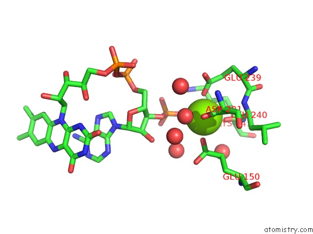
Mono view
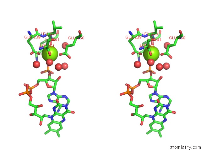
Stereo pair view
Mono view
Stereo pair view
A full contact list of Magnesium with other atoms in the Mg binding
site number 1 of Crystal Structure of S. Pombe RAI1 in Complex with 3'-Fadp within 5.0Å range:
|
Magnesium binding site 2 out of 2 in 6wug
Go back to
Magnesium binding site 2 out
of 2 in the Crystal Structure of S. Pombe RAI1 in Complex with 3'-Fadp
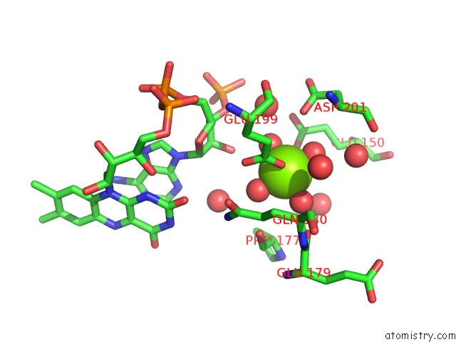
Mono view
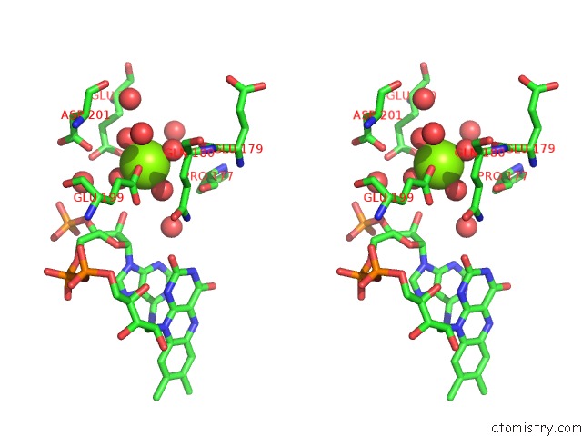
Stereo pair view
Mono view
Stereo pair view
A full contact list of Magnesium with other atoms in the Mg binding
site number 2 of Crystal Structure of S. Pombe RAI1 in Complex with 3'-Fadp within 5.0Å range:
|
Reference:
S.K.Doamekpor,
E.Grudzien-Nogalska,
A.Mlynarska-Cieslak,
J.Kowalska,
M.Kiledjian,
L.Tong.
Dxo/RAI1 Enzymes Remove 5'-End Fad and Dephospho-Coa Caps on Rnas. Nucleic Acids Res. 2020.
ISSN: ESSN 1362-4962
PubMed: 32374864
DOI: 10.1093/NAR/GKAA297
Page generated: Tue Oct 1 23:14:41 2024
ISSN: ESSN 1362-4962
PubMed: 32374864
DOI: 10.1093/NAR/GKAA297
Last articles
Zn in 9MJ5Zn in 9HNW
Zn in 9G0L
Zn in 9FNE
Zn in 9DZN
Zn in 9E0I
Zn in 9D32
Zn in 9DAK
Zn in 8ZXC
Zn in 8ZUF