Magnesium »
PDB 6wwn-6x4b »
6x1g »
Magnesium in PDB 6x1g: Crystal Structure of A Gef Domain From the Orientia Tsutsugamushi Protein Otdub in Complex with RAC1
Enzymatic activity of Crystal Structure of A Gef Domain From the Orientia Tsutsugamushi Protein Otdub in Complex with RAC1
All present enzymatic activity of Crystal Structure of A Gef Domain From the Orientia Tsutsugamushi Protein Otdub in Complex with RAC1:
3.6.5.2;
3.6.5.2;
Protein crystallography data
The structure of Crystal Structure of A Gef Domain From the Orientia Tsutsugamushi Protein Otdub in Complex with RAC1, PDB code: 6x1g
was solved by
C.S.Lim,
Y.Xiong,
with X-Ray Crystallography technique. A brief refinement statistics is given in the table below:
| Resolution Low / High (Å) | 48.05 / 1.60 |
| Space group | P 1 |
| Cell size a, b, c (Å), α, β, γ (°) | 50.063, 54.160, 94.004, 83.37, 76.34, 62.52 |
| R / Rfree (%) | 18.1 / 21.8 |
Magnesium Binding Sites:
The binding sites of Magnesium atom in the Crystal Structure of A Gef Domain From the Orientia Tsutsugamushi Protein Otdub in Complex with RAC1
(pdb code 6x1g). This binding sites where shown within
5.0 Angstroms radius around Magnesium atom.
In total only one binding site of Magnesium was determined in the Crystal Structure of A Gef Domain From the Orientia Tsutsugamushi Protein Otdub in Complex with RAC1, PDB code: 6x1g:
In total only one binding site of Magnesium was determined in the Crystal Structure of A Gef Domain From the Orientia Tsutsugamushi Protein Otdub in Complex with RAC1, PDB code: 6x1g:
Magnesium binding site 1 out of 1 in 6x1g
Go back to
Magnesium binding site 1 out
of 1 in the Crystal Structure of A Gef Domain From the Orientia Tsutsugamushi Protein Otdub in Complex with RAC1
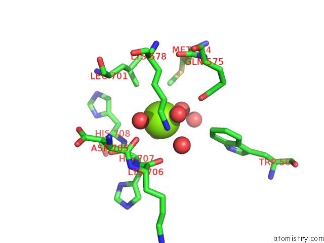
Mono view
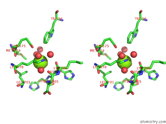
Stereo pair view
Mono view
Stereo pair view
A full contact list of Magnesium with other atoms in the Mg binding
site number 1 of Crystal Structure of A Gef Domain From the Orientia Tsutsugamushi Protein Otdub in Complex with RAC1 within 5.0Å range:
|
Reference:
C.Lim,
J.M.Berk,
A.Blaise,
J.Bircher,
A.J.Koleske,
M.Hochstrasser,
Y.Xiong.
Crystal Structure of A Guanine Nucleotide Exchange Factor Encoded By the Scrub Typhus Pathogen Orientia Tsutsugamushi . Proc.Natl.Acad.Sci.Usa 2020.
ISSN: ESSN 1091-6490
PubMed: 33184172
DOI: 10.1073/PNAS.2018163117
Page generated: Tue Oct 1 23:20:32 2024
ISSN: ESSN 1091-6490
PubMed: 33184172
DOI: 10.1073/PNAS.2018163117
Last articles
Zn in 9MJ5Zn in 9HNW
Zn in 9G0L
Zn in 9FNE
Zn in 9DZN
Zn in 9E0I
Zn in 9D32
Zn in 9DAK
Zn in 8ZXC
Zn in 8ZUF