Magnesium »
PDB 6xg6-6xmq »
6xln »
Magnesium in PDB 6xln: Cryo-Em Structure of E. Coli Rnap-Dna Elongation Complex 2 (RDE2) in Ecmrr-Dependent Transcription
Enzymatic activity of Cryo-Em Structure of E. Coli Rnap-Dna Elongation Complex 2 (RDE2) in Ecmrr-Dependent Transcription
All present enzymatic activity of Cryo-Em Structure of E. Coli Rnap-Dna Elongation Complex 2 (RDE2) in Ecmrr-Dependent Transcription:
2.7.7.6;
2.7.7.6;
Other elements in 6xln:
The structure of Cryo-Em Structure of E. Coli Rnap-Dna Elongation Complex 2 (RDE2) in Ecmrr-Dependent Transcription also contains other interesting chemical elements:
| Zinc | (Zn) | 2 atoms |
Magnesium Binding Sites:
The binding sites of Magnesium atom in the Cryo-Em Structure of E. Coli Rnap-Dna Elongation Complex 2 (RDE2) in Ecmrr-Dependent Transcription
(pdb code 6xln). This binding sites where shown within
5.0 Angstroms radius around Magnesium atom.
In total only one binding site of Magnesium was determined in the Cryo-Em Structure of E. Coli Rnap-Dna Elongation Complex 2 (RDE2) in Ecmrr-Dependent Transcription, PDB code: 6xln:
In total only one binding site of Magnesium was determined in the Cryo-Em Structure of E. Coli Rnap-Dna Elongation Complex 2 (RDE2) in Ecmrr-Dependent Transcription, PDB code: 6xln:
Magnesium binding site 1 out of 1 in 6xln
Go back to
Magnesium binding site 1 out
of 1 in the Cryo-Em Structure of E. Coli Rnap-Dna Elongation Complex 2 (RDE2) in Ecmrr-Dependent Transcription
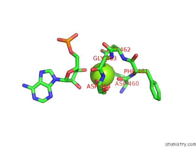
Mono view
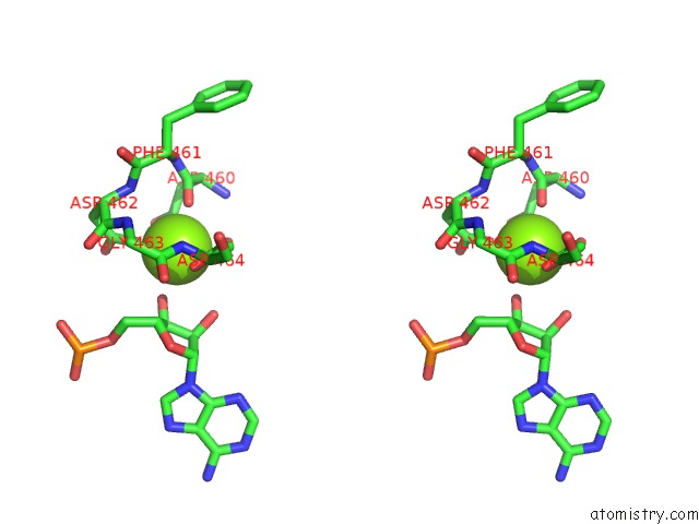
Stereo pair view
Mono view
Stereo pair view
A full contact list of Magnesium with other atoms in the Mg binding
site number 1 of Cryo-Em Structure of E. Coli Rnap-Dna Elongation Complex 2 (RDE2) in Ecmrr-Dependent Transcription within 5.0Å range:
|
Reference:
Y.Yang,
C.Liu,
W.Shi,
D.G.Schatz,
Y.Hu,
B.Liu.
Structural Visualization of Bacterial Multidrug-Activated Transcription To Be Published.
Page generated: Tue Oct 1 23:35:44 2024
Last articles
Zn in 9J0NZn in 9J0O
Zn in 9J0P
Zn in 9FJX
Zn in 9EKB
Zn in 9C0F
Zn in 9CAH
Zn in 9CH0
Zn in 9CH3
Zn in 9CH1