Magnesium »
PDB 6y0t-6ya8 »
6y5y »
Magnesium in PDB 6y5y: Structure of New Jersey Polyomavirus VP1 in Complex with 3'- Sialyllactose
Protein crystallography data
The structure of Structure of New Jersey Polyomavirus VP1 in Complex with 3'- Sialyllactose, PDB code: 6y5y
was solved by
L.J.Stroh,
N.H.Rustmeier,
T.Stehle,
with X-Ray Crystallography technique. A brief refinement statistics is given in the table below:
| Resolution Low / High (Å) | 48.20 / 1.80 |
| Space group | P 1 21 1 |
| Cell size a, b, c (Å), α, β, γ (°) | 86.381, 151.082, 130.617, 90.00, 106.56, 90.00 |
| R / Rfree (%) | 16.6 / 20.6 |
Magnesium Binding Sites:
The binding sites of Magnesium atom in the Structure of New Jersey Polyomavirus VP1 in Complex with 3'- Sialyllactose
(pdb code 6y5y). This binding sites where shown within
5.0 Angstroms radius around Magnesium atom.
In total 10 binding sites of Magnesium where determined in the Structure of New Jersey Polyomavirus VP1 in Complex with 3'- Sialyllactose, PDB code: 6y5y:
Jump to Magnesium binding site number: 1; 2; 3; 4; 5; 6; 7; 8; 9; 10;
In total 10 binding sites of Magnesium where determined in the Structure of New Jersey Polyomavirus VP1 in Complex with 3'- Sialyllactose, PDB code: 6y5y:
Jump to Magnesium binding site number: 1; 2; 3; 4; 5; 6; 7; 8; 9; 10;
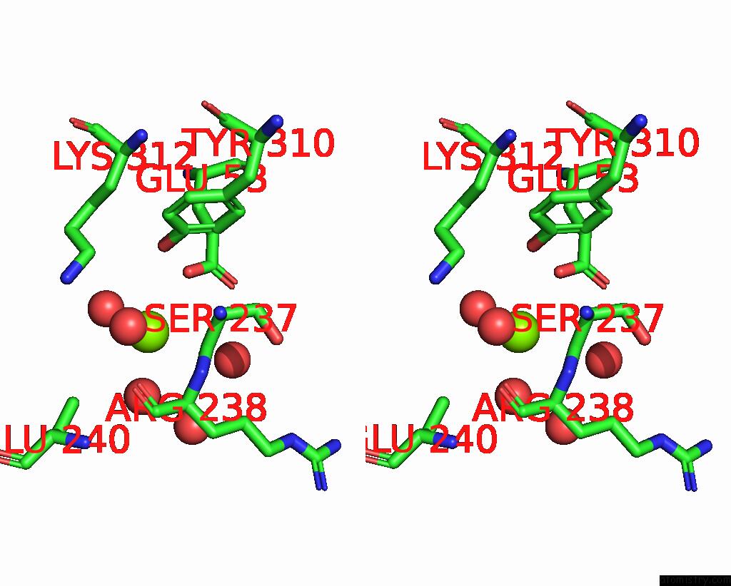
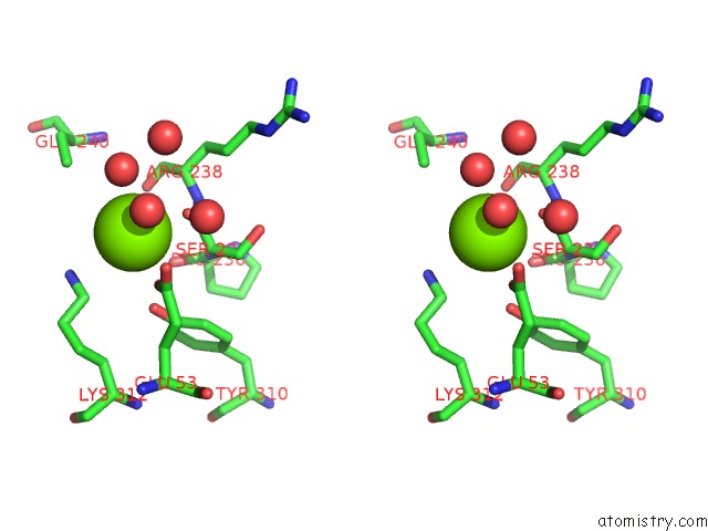
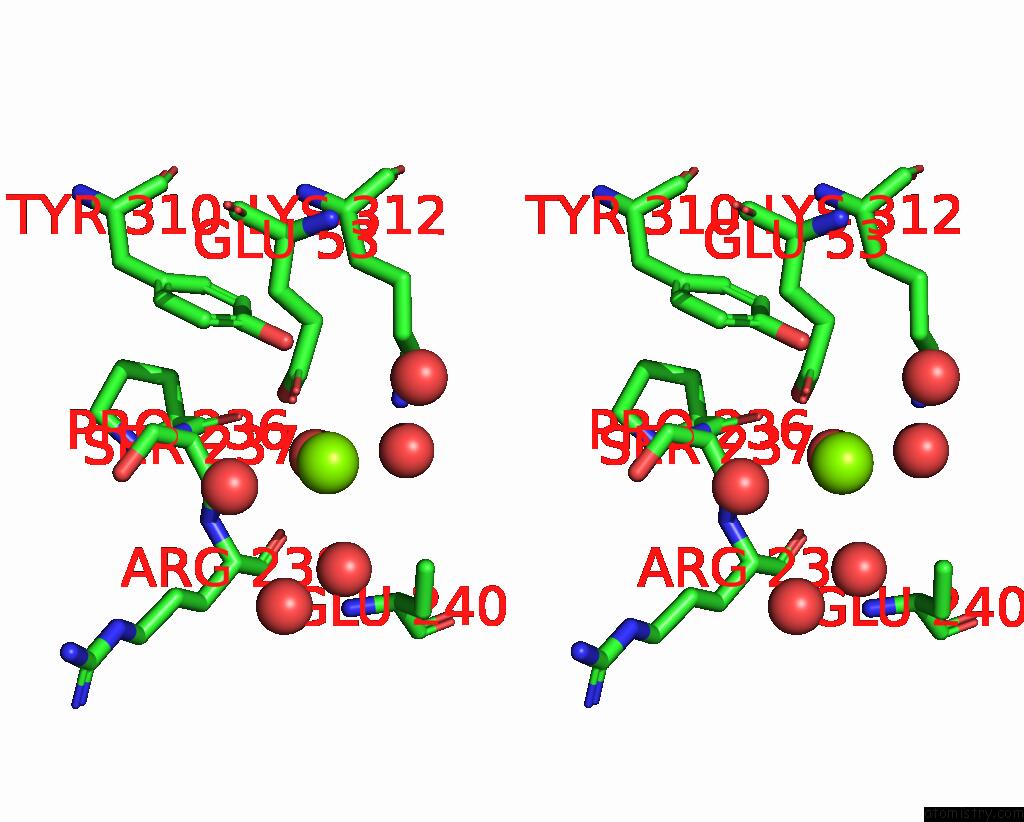
Magnesium binding site 1 out of 10 in 6y5y
Go back to
Magnesium binding site 1 out
of 10 in the Structure of New Jersey Polyomavirus VP1 in Complex with 3'- Sialyllactose
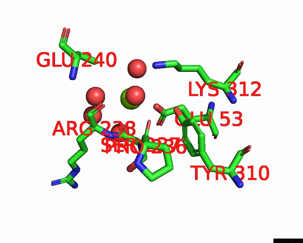
Mono view
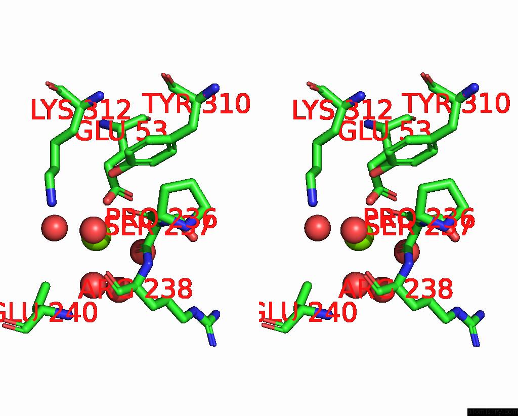
Stereo pair view
Mono view
Stereo pair view
A full contact list of Magnesium with other atoms in the Mg binding
site number 1 of Structure of New Jersey Polyomavirus VP1 in Complex with 3'- Sialyllactose within 5.0Å range:
|
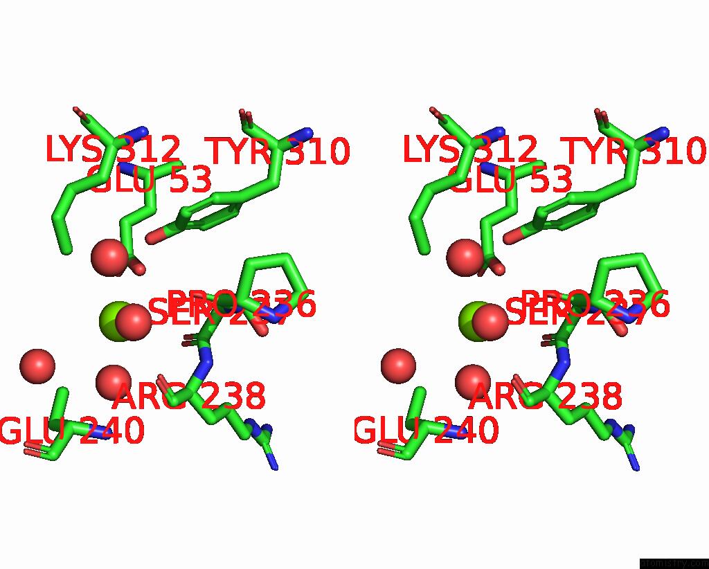
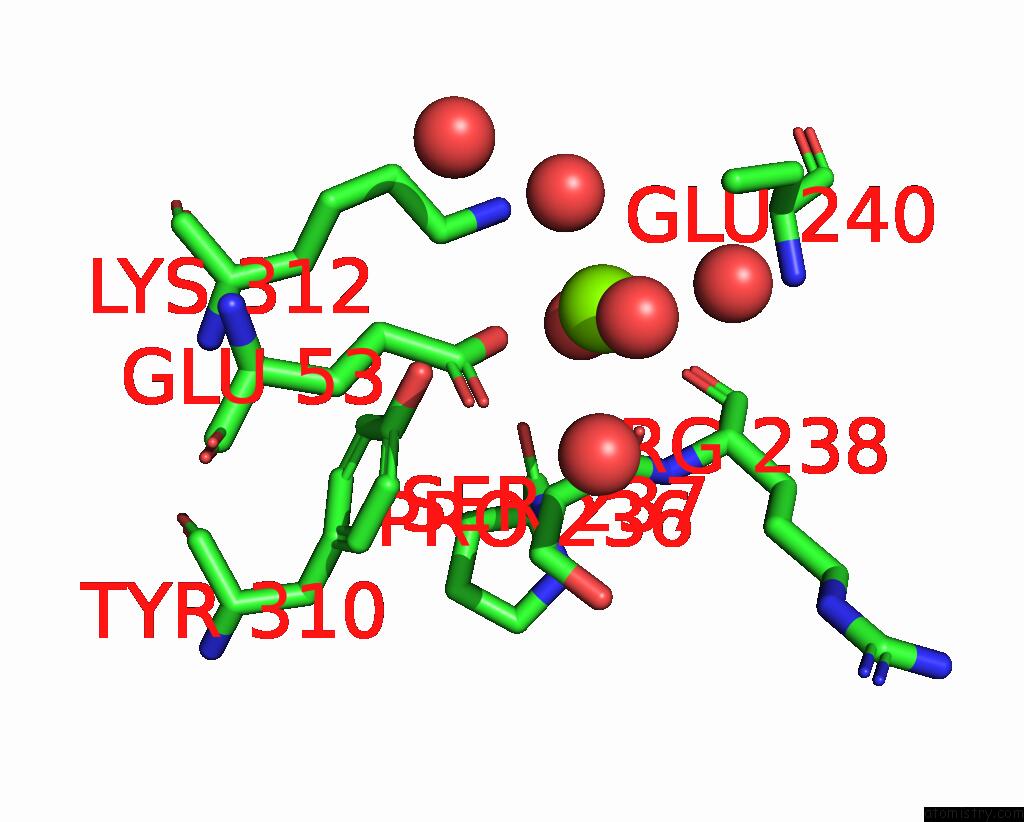
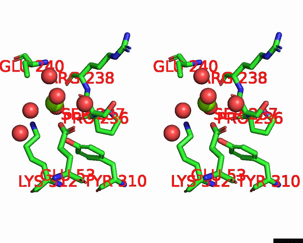
Magnesium binding site 2 out of 10 in 6y5y
Go back to
Magnesium binding site 2 out
of 10 in the Structure of New Jersey Polyomavirus VP1 in Complex with 3'- Sialyllactose
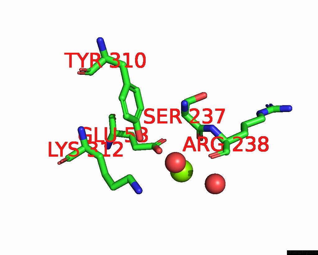
Mono view

Stereo pair view
Mono view
Stereo pair view
A full contact list of Magnesium with other atoms in the Mg binding
site number 2 of Structure of New Jersey Polyomavirus VP1 in Complex with 3'- Sialyllactose within 5.0Å range:
|
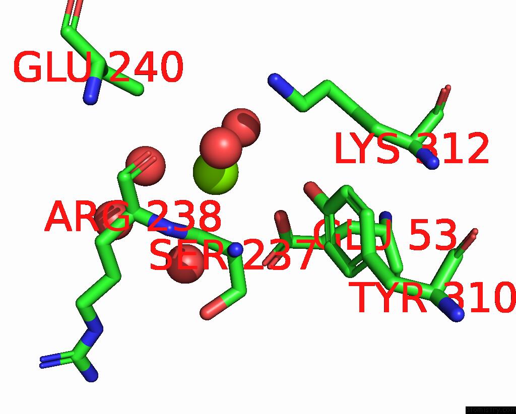
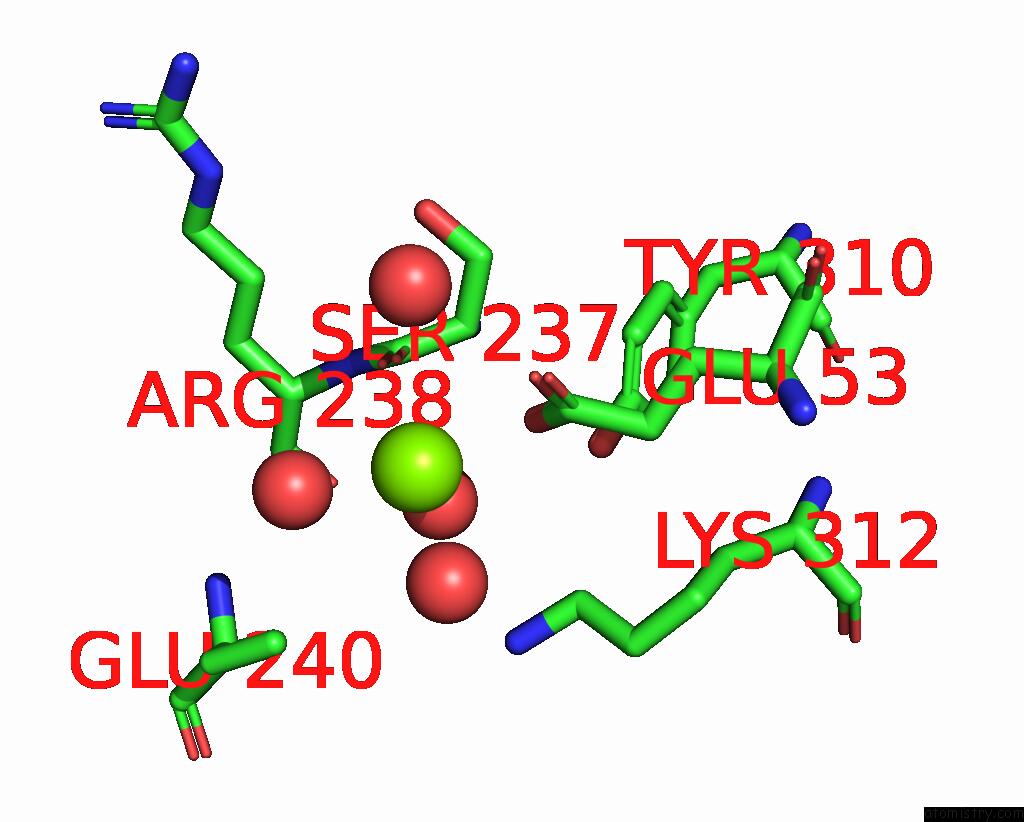
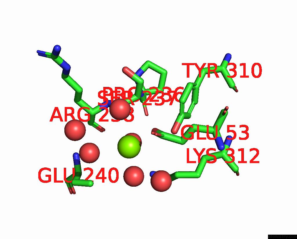
Magnesium binding site 3 out of 10 in 6y5y
Go back to
Magnesium binding site 3 out
of 10 in the Structure of New Jersey Polyomavirus VP1 in Complex with 3'- Sialyllactose

Mono view

Stereo pair view
Mono view
Stereo pair view
A full contact list of Magnesium with other atoms in the Mg binding
site number 3 of Structure of New Jersey Polyomavirus VP1 in Complex with 3'- Sialyllactose within 5.0Å range:
|
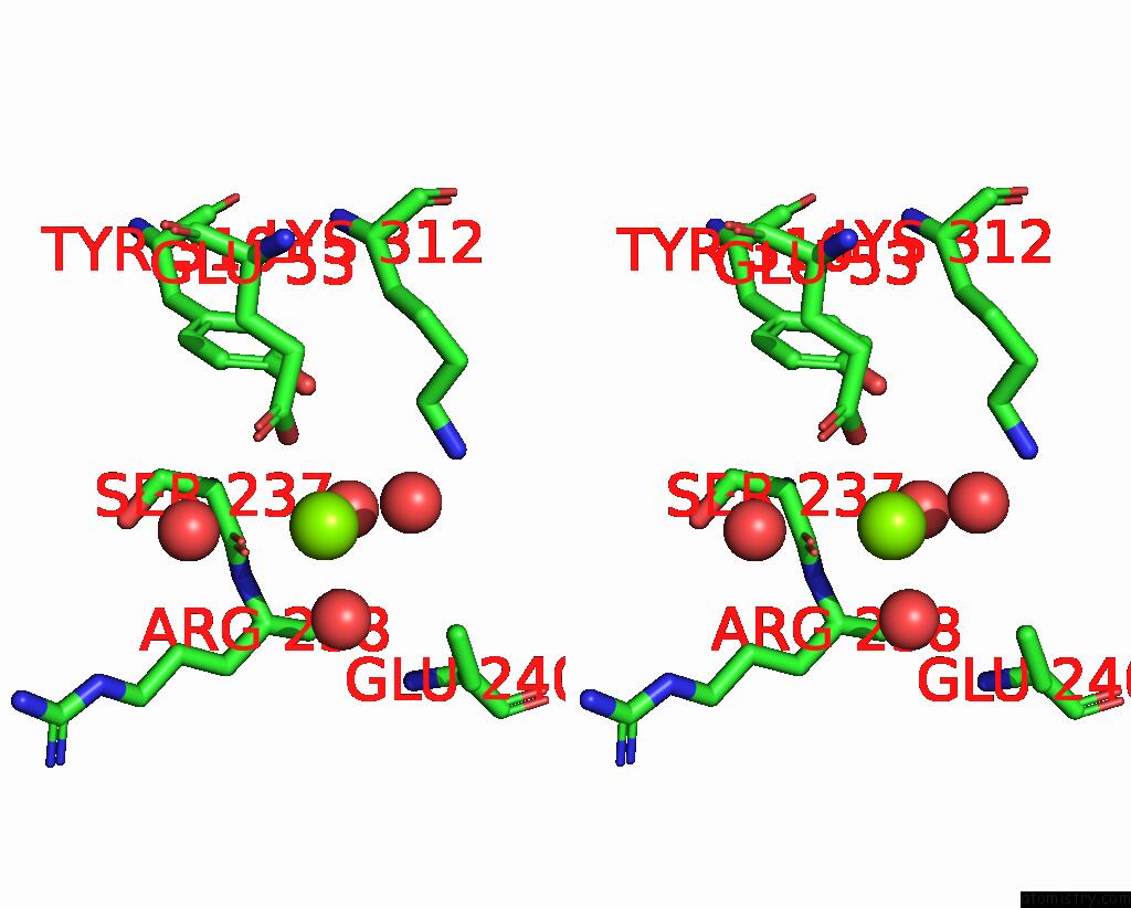
Magnesium binding site 4 out of 10 in 6y5y
Go back to
Magnesium binding site 4 out
of 10 in the Structure of New Jersey Polyomavirus VP1 in Complex with 3'- Sialyllactose
Mono view
Stereo pair view
Mono view
Stereo pair view
A full contact list of Magnesium with other atoms in the Mg binding
site number 4 of Structure of New Jersey Polyomavirus VP1 in Complex with 3'- Sialyllactose within 5.0Å range:
|
Magnesium binding site 5 out of 10 in 6y5y
Go back to
Magnesium binding site 5 out
of 10 in the Structure of New Jersey Polyomavirus VP1 in Complex with 3'- Sialyllactose

Mono view
Stereo pair view
Mono view
Stereo pair view
A full contact list of Magnesium with other atoms in the Mg binding
site number 5 of Structure of New Jersey Polyomavirus VP1 in Complex with 3'- Sialyllactose within 5.0Å range:
|
Magnesium binding site 6 out of 10 in 6y5y
Go back to
Magnesium binding site 6 out
of 10 in the Structure of New Jersey Polyomavirus VP1 in Complex with 3'- Sialyllactose
Mono view
Stereo pair view
Mono view
Stereo pair view
A full contact list of Magnesium with other atoms in the Mg binding
site number 6 of Structure of New Jersey Polyomavirus VP1 in Complex with 3'- Sialyllactose within 5.0Å range:
|
Magnesium binding site 7 out of 10 in 6y5y
Go back to
Magnesium binding site 7 out
of 10 in the Structure of New Jersey Polyomavirus VP1 in Complex with 3'- Sialyllactose

Mono view
Stereo pair view
Mono view
Stereo pair view
A full contact list of Magnesium with other atoms in the Mg binding
site number 7 of Structure of New Jersey Polyomavirus VP1 in Complex with 3'- Sialyllactose within 5.0Å range:
|
Magnesium binding site 8 out of 10 in 6y5y
Go back to
Magnesium binding site 8 out
of 10 in the Structure of New Jersey Polyomavirus VP1 in Complex with 3'- Sialyllactose
Mono view
Stereo pair view
Mono view
Stereo pair view
A full contact list of Magnesium with other atoms in the Mg binding
site number 8 of Structure of New Jersey Polyomavirus VP1 in Complex with 3'- Sialyllactose within 5.0Å range:
|
Magnesium binding site 9 out of 10 in 6y5y
Go back to
Magnesium binding site 9 out
of 10 in the Structure of New Jersey Polyomavirus VP1 in Complex with 3'- Sialyllactose
Mono view
Stereo pair view
Mono view
Stereo pair view
A full contact list of Magnesium with other atoms in the Mg binding
site number 9 of Structure of New Jersey Polyomavirus VP1 in Complex with 3'- Sialyllactose within 5.0Å range:
|
Magnesium binding site 10 out of 10 in 6y5y
Go back to
Magnesium binding site 10 out
of 10 in the Structure of New Jersey Polyomavirus VP1 in Complex with 3'- Sialyllactose

Mono view

Stereo pair view
Mono view
Stereo pair view
A full contact list of Magnesium with other atoms in the Mg binding
site number 10 of Structure of New Jersey Polyomavirus VP1 in Complex with 3'- Sialyllactose within 5.0Å range:
|
Reference:
L.J.Stroh,
N.H.Rustmeier,
B.S.Blaum,
J.Botsch,
P.Rossler,
F.Wedekink,
W.I.Lipkin,
N.Mishra,
T.Stehle.
Structural Basis and Evolution of Glycan Receptor Specificities Within the Polyomavirus Family. Mbio V. 11 2020.
ISSN: ESSN 2150-7511
PubMed: 32723915
DOI: 10.1128/MBIO.00745-20
Page generated: Wed Oct 2 00:10:28 2024
ISSN: ESSN 2150-7511
PubMed: 32723915
DOI: 10.1128/MBIO.00745-20
Last articles
Zn in 9J0NZn in 9J0O
Zn in 9J0P
Zn in 9FJX
Zn in 9EKB
Zn in 9C0F
Zn in 9CAH
Zn in 9CH0
Zn in 9CH3
Zn in 9CH1