Magnesium »
PDB 6yac-6ykt »
6ydm »
Magnesium in PDB 6ydm: Beta-Phosphoglucomutase From Lactococcus Lactis with Citrate, Tris and Acetate Bound
Enzymatic activity of Beta-Phosphoglucomutase From Lactococcus Lactis with Citrate, Tris and Acetate Bound
All present enzymatic activity of Beta-Phosphoglucomutase From Lactococcus Lactis with Citrate, Tris and Acetate Bound:
5.4.2.6;
5.4.2.6;
Protein crystallography data
The structure of Beta-Phosphoglucomutase From Lactococcus Lactis with Citrate, Tris and Acetate Bound, PDB code: 6ydm
was solved by
H.P.Wood,
F.A.Cruz-Navarrete,
N.J.Baxter,
C.R.Trevitt,
A.J.Robertson,
S.R.Dix,
A.M.Hounslow,
M.J.Cliff,
J.P.Waltho,
with X-Ray Crystallography technique. A brief refinement statistics is given in the table below:
| Resolution Low / High (Å) | 46.61 / 2.10 |
| Space group | P 21 21 21 |
| Cell size a, b, c (Å), α, β, γ (°) | 53.130, 76.640, 117.260, 90.00, 90.00, 90.00 |
| R / Rfree (%) | 23 / 29 |
Magnesium Binding Sites:
The binding sites of Magnesium atom in the Beta-Phosphoglucomutase From Lactococcus Lactis with Citrate, Tris and Acetate Bound
(pdb code 6ydm). This binding sites where shown within
5.0 Angstroms radius around Magnesium atom.
In total 2 binding sites of Magnesium where determined in the Beta-Phosphoglucomutase From Lactococcus Lactis with Citrate, Tris and Acetate Bound, PDB code: 6ydm:
Jump to Magnesium binding site number: 1; 2;
In total 2 binding sites of Magnesium where determined in the Beta-Phosphoglucomutase From Lactococcus Lactis with Citrate, Tris and Acetate Bound, PDB code: 6ydm:
Jump to Magnesium binding site number: 1; 2;
Magnesium binding site 1 out of 2 in 6ydm
Go back to
Magnesium binding site 1 out
of 2 in the Beta-Phosphoglucomutase From Lactococcus Lactis with Citrate, Tris and Acetate Bound
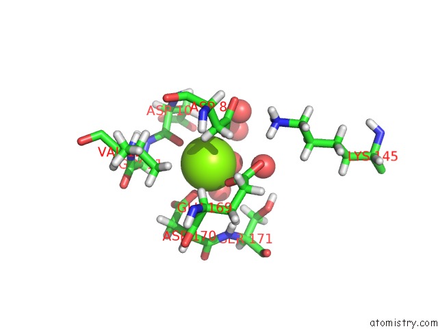
Mono view
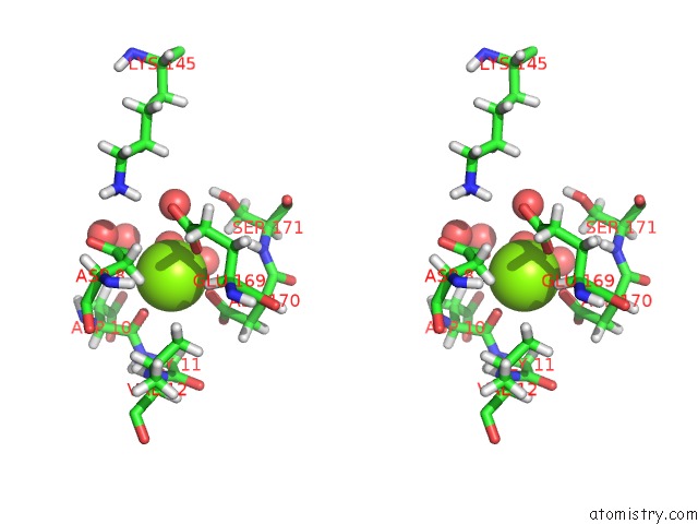
Stereo pair view
Mono view
Stereo pair view
A full contact list of Magnesium with other atoms in the Mg binding
site number 1 of Beta-Phosphoglucomutase From Lactococcus Lactis with Citrate, Tris and Acetate Bound within 5.0Å range:
|
Magnesium binding site 2 out of 2 in 6ydm
Go back to
Magnesium binding site 2 out
of 2 in the Beta-Phosphoglucomutase From Lactococcus Lactis with Citrate, Tris and Acetate Bound
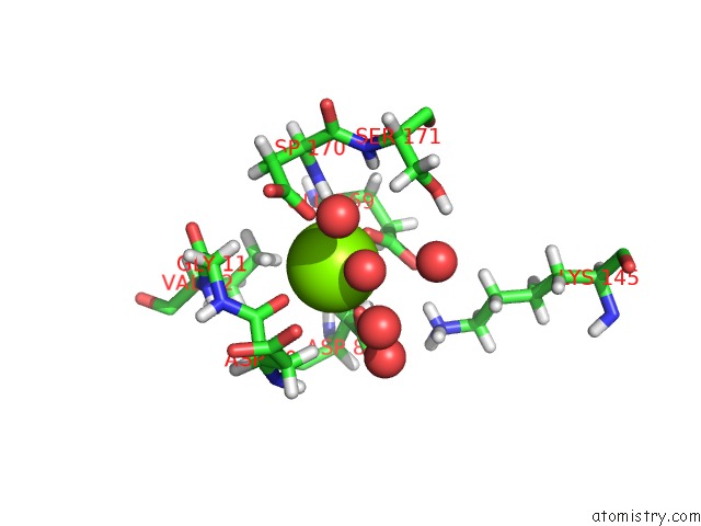
Mono view
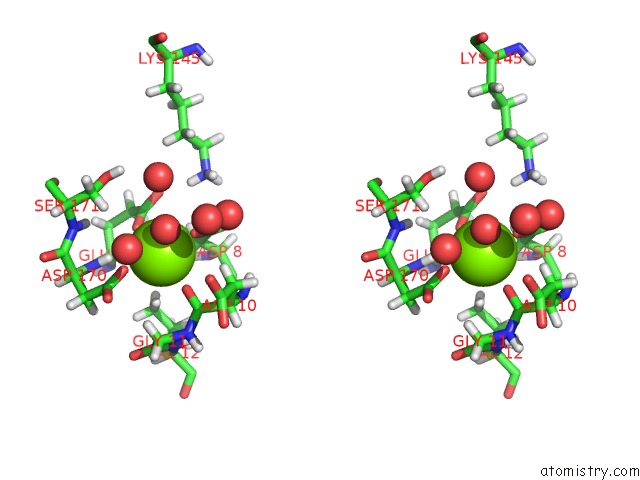
Stereo pair view
Mono view
Stereo pair view
A full contact list of Magnesium with other atoms in the Mg binding
site number 2 of Beta-Phosphoglucomutase From Lactococcus Lactis with Citrate, Tris and Acetate Bound within 5.0Å range:
|
Reference:
H.P.Wood,
F.A.Cruz-Navarrete,
N.J.Baxter,
C.R.Trevitt,
A.J.Robertson,
S.R.Dix,
A.M.Hounslow,
M.J.Cliff,
J.P.Waltho.
Allomorphy As A Mechanism of Post-Translational Control of Enzyme Activity. Nat Commun V. 11 5538 2020.
ISSN: ESSN 2041-1723
PubMed: 33139716
DOI: 10.1038/S41467-020-19215-9
Page generated: Wed Oct 2 00:18:27 2024
ISSN: ESSN 2041-1723
PubMed: 33139716
DOI: 10.1038/S41467-020-19215-9
Last articles
Zn in 9J0NZn in 9J0O
Zn in 9J0P
Zn in 9FJX
Zn in 9EKB
Zn in 9C0F
Zn in 9CAH
Zn in 9CH0
Zn in 9CH3
Zn in 9CH1