Magnesium »
PDB 6ykv-6yxt »
6yp4 »
Magnesium in PDB 6yp4: Putative Adenylyl Cyclase HPAC1 From Hippeastrum Reveals A Dominant Triphophatase Activity
Protein crystallography data
The structure of Putative Adenylyl Cyclase HPAC1 From Hippeastrum Reveals A Dominant Triphophatase Activity, PDB code: 6yp4
was solved by
S.Kleinboelting,
C.Steegborn,
with X-Ray Crystallography technique. A brief refinement statistics is given in the table below:
| Resolution Low / High (Å) | 46.39 / 1.95 |
| Space group | P 41 21 2 |
| Cell size a, b, c (Å), α, β, γ (°) | 59.808, 59.808, 146.982, 90.00, 90.00, 90.00 |
| R / Rfree (%) | 19.5 / 25.2 |
Other elements in 6yp4:
The structure of Putative Adenylyl Cyclase HPAC1 From Hippeastrum Reveals A Dominant Triphophatase Activity also contains other interesting chemical elements:
| Chlorine | (Cl) | 3 atoms |
Magnesium Binding Sites:
The binding sites of Magnesium atom in the Putative Adenylyl Cyclase HPAC1 From Hippeastrum Reveals A Dominant Triphophatase Activity
(pdb code 6yp4). This binding sites where shown within
5.0 Angstroms radius around Magnesium atom.
In total 2 binding sites of Magnesium where determined in the Putative Adenylyl Cyclase HPAC1 From Hippeastrum Reveals A Dominant Triphophatase Activity, PDB code: 6yp4:
Jump to Magnesium binding site number: 1; 2;
In total 2 binding sites of Magnesium where determined in the Putative Adenylyl Cyclase HPAC1 From Hippeastrum Reveals A Dominant Triphophatase Activity, PDB code: 6yp4:
Jump to Magnesium binding site number: 1; 2;
Magnesium binding site 1 out of 2 in 6yp4
Go back to
Magnesium binding site 1 out
of 2 in the Putative Adenylyl Cyclase HPAC1 From Hippeastrum Reveals A Dominant Triphophatase Activity
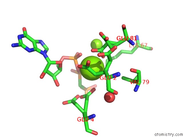
Mono view
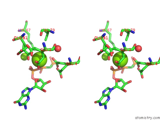
Stereo pair view
Mono view
Stereo pair view
A full contact list of Magnesium with other atoms in the Mg binding
site number 1 of Putative Adenylyl Cyclase HPAC1 From Hippeastrum Reveals A Dominant Triphophatase Activity within 5.0Å range:
|
Magnesium binding site 2 out of 2 in 6yp4
Go back to
Magnesium binding site 2 out
of 2 in the Putative Adenylyl Cyclase HPAC1 From Hippeastrum Reveals A Dominant Triphophatase Activity
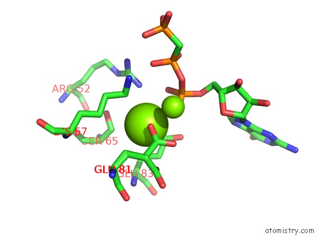
Mono view
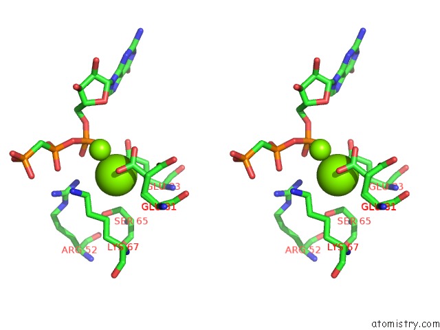
Stereo pair view
Mono view
Stereo pair view
A full contact list of Magnesium with other atoms in the Mg binding
site number 2 of Putative Adenylyl Cyclase HPAC1 From Hippeastrum Reveals A Dominant Triphophatase Activity within 5.0Å range:
|
Reference:
S.Kleinboelting,
J.Miehling,
C.Steegborn.
Crystal Structure and Enzymatic Characterization of the Putative Adenylyl Cyclase HPAC1 From Hippeastrum Reveal Dominant Triphosphatase Activity. J.Struct.Biol. V. 212 07649 2020.
ISSN: ESSN 1095-8657
PubMed: 33075486
DOI: 10.1016/J.JSB.2020.107649
Page generated: Wed Oct 2 01:03:27 2024
ISSN: ESSN 1095-8657
PubMed: 33075486
DOI: 10.1016/J.JSB.2020.107649
Last articles
Zn in 9MJ5Zn in 9HNW
Zn in 9G0L
Zn in 9FNE
Zn in 9DZN
Zn in 9E0I
Zn in 9D32
Zn in 9DAK
Zn in 8ZXC
Zn in 8ZUF