Magnesium »
PDB 6ykv-6yxt »
6ypu »
Magnesium in PDB 6ypu: Acinetobacter Baumannii Ribosome-Amikacin Complex - 30S Subunit Body
Magnesium Binding Sites:
Pages:
>>> Page 1 <<< Page 2, Binding sites: 11 - 20; Page 3, Binding sites: 21 - 30; Page 4, Binding sites: 31 - 40; Page 5, Binding sites: 41 - 50; Page 6, Binding sites: 51 - 59;Binding sites:
The binding sites of Magnesium atom in the Acinetobacter Baumannii Ribosome-Amikacin Complex - 30S Subunit Body (pdb code 6ypu). This binding sites where shown within 5.0 Angstroms radius around Magnesium atom.In total 59 binding sites of Magnesium where determined in the Acinetobacter Baumannii Ribosome-Amikacin Complex - 30S Subunit Body, PDB code: 6ypu:
Jump to Magnesium binding site number: 1; 2; 3; 4; 5; 6; 7; 8; 9; 10;
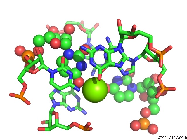
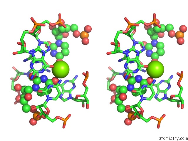
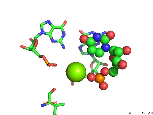
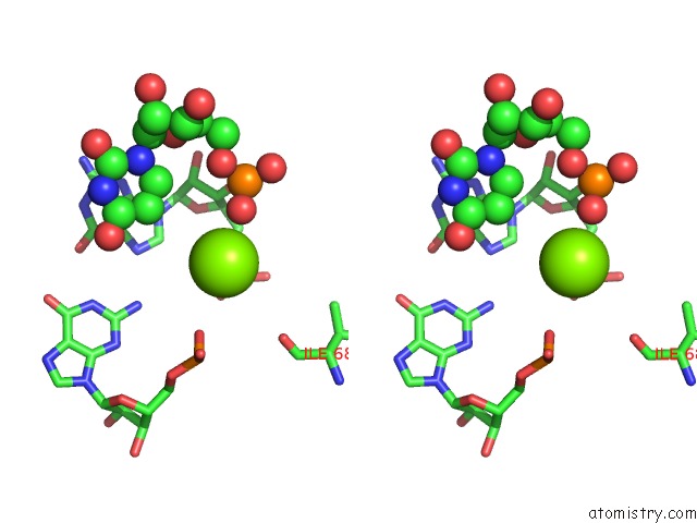
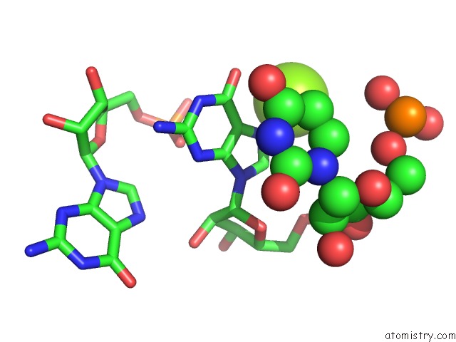
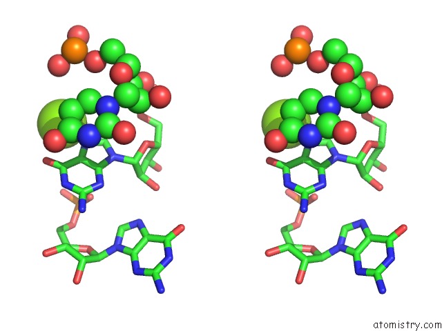
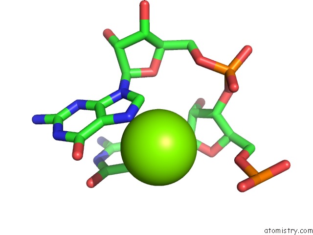
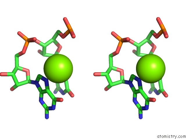
Magnesium binding site 1 out of 59 in 6ypu
Go back to
Magnesium binding site 1 out
of 59 in the Acinetobacter Baumannii Ribosome-Amikacin Complex - 30S Subunit Body
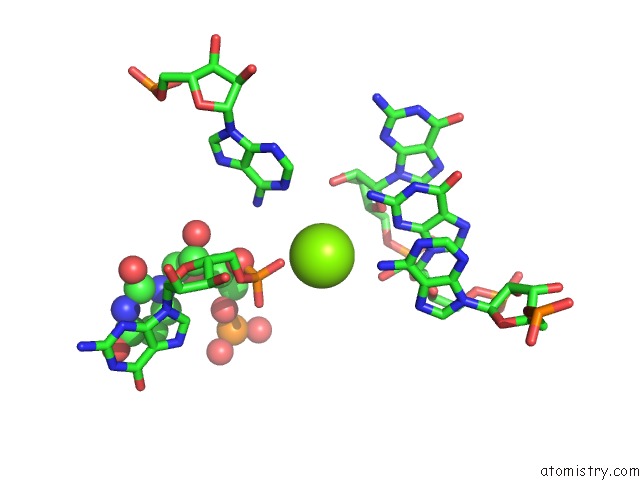
Mono view
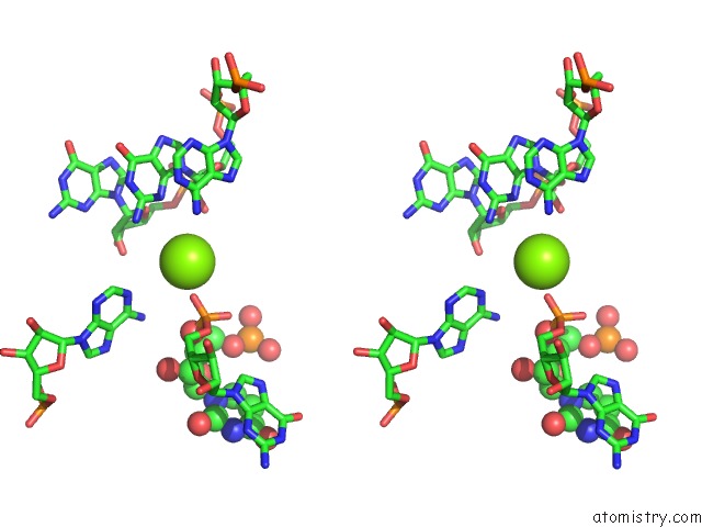
Stereo pair view
Mono view
Stereo pair view
A full contact list of Magnesium with other atoms in the Mg binding
site number 1 of Acinetobacter Baumannii Ribosome-Amikacin Complex - 30S Subunit Body within 5.0Å range:
|
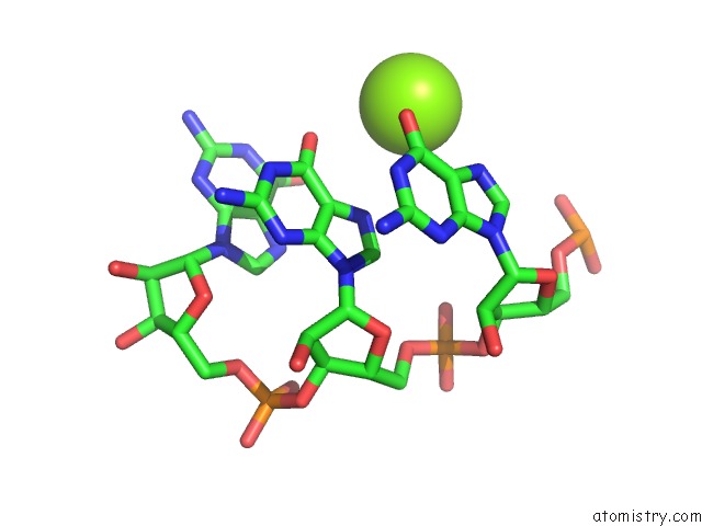
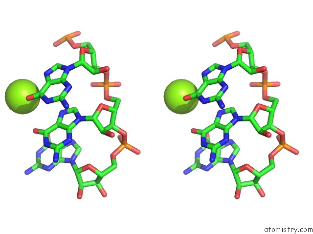
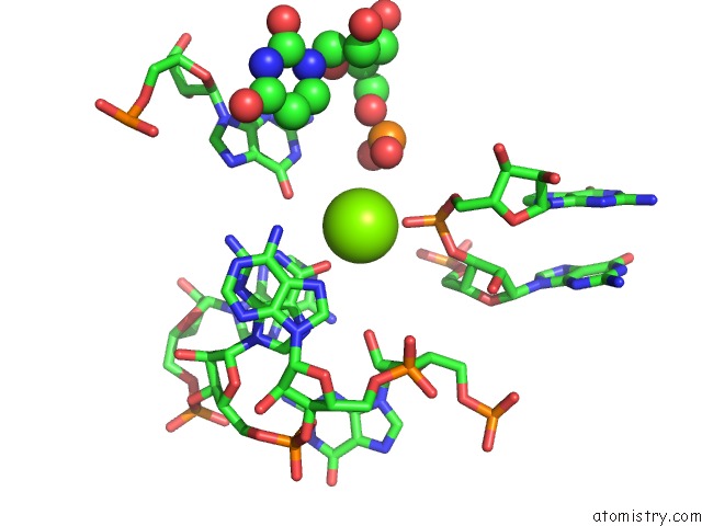
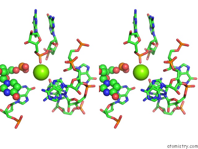
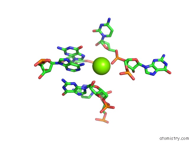
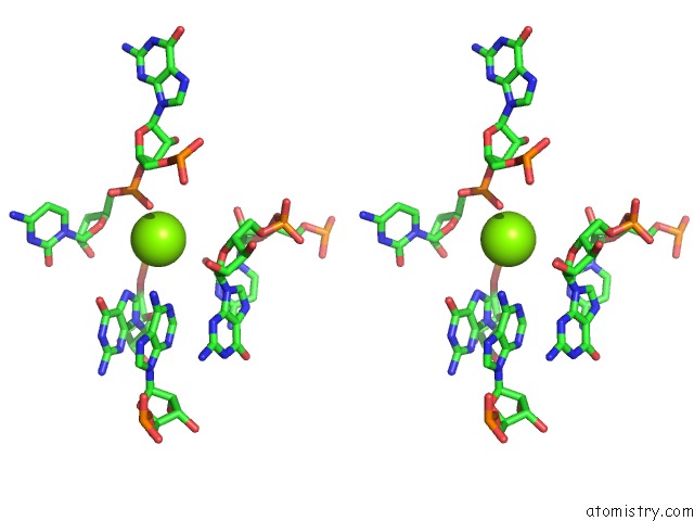
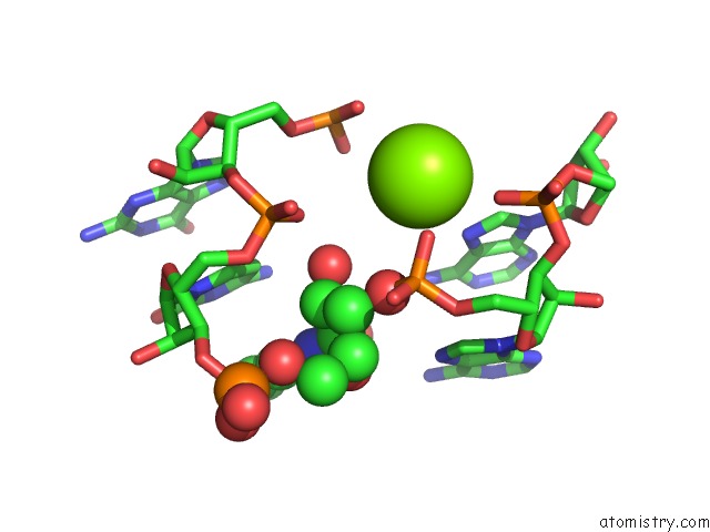
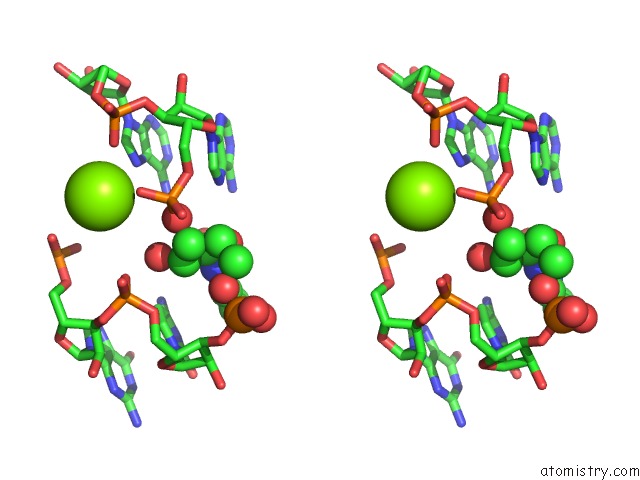
Magnesium binding site 2 out of 59 in 6ypu
Go back to
Magnesium binding site 2 out
of 59 in the Acinetobacter Baumannii Ribosome-Amikacin Complex - 30S Subunit Body
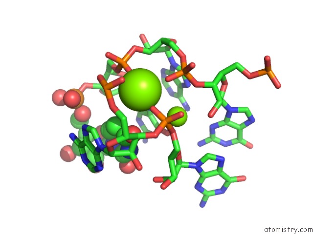
Mono view
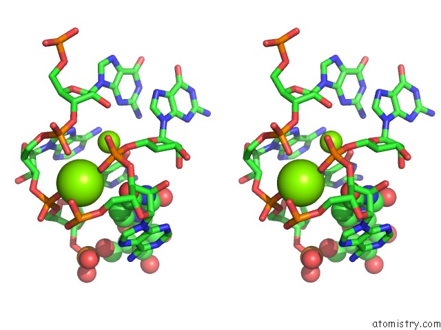
Stereo pair view
Mono view
Stereo pair view
A full contact list of Magnesium with other atoms in the Mg binding
site number 2 of Acinetobacter Baumannii Ribosome-Amikacin Complex - 30S Subunit Body within 5.0Å range:
|
Magnesium binding site 3 out of 59 in 6ypu
Go back to
Magnesium binding site 3 out
of 59 in the Acinetobacter Baumannii Ribosome-Amikacin Complex - 30S Subunit Body
Mono view
Stereo pair view
Mono view
Stereo pair view
A full contact list of Magnesium with other atoms in the Mg binding
site number 3 of Acinetobacter Baumannii Ribosome-Amikacin Complex - 30S Subunit Body within 5.0Å range:
|
Magnesium binding site 4 out of 59 in 6ypu
Go back to
Magnesium binding site 4 out
of 59 in the Acinetobacter Baumannii Ribosome-Amikacin Complex - 30S Subunit Body
Mono view
Stereo pair view
Mono view
Stereo pair view
A full contact list of Magnesium with other atoms in the Mg binding
site number 4 of Acinetobacter Baumannii Ribosome-Amikacin Complex - 30S Subunit Body within 5.0Å range:
|
Magnesium binding site 5 out of 59 in 6ypu
Go back to
Magnesium binding site 5 out
of 59 in the Acinetobacter Baumannii Ribosome-Amikacin Complex - 30S Subunit Body
Mono view
Stereo pair view
Mono view
Stereo pair view
A full contact list of Magnesium with other atoms in the Mg binding
site number 5 of Acinetobacter Baumannii Ribosome-Amikacin Complex - 30S Subunit Body within 5.0Å range:
|
Magnesium binding site 6 out of 59 in 6ypu
Go back to
Magnesium binding site 6 out
of 59 in the Acinetobacter Baumannii Ribosome-Amikacin Complex - 30S Subunit Body
Mono view
Stereo pair view
Mono view
Stereo pair view
A full contact list of Magnesium with other atoms in the Mg binding
site number 6 of Acinetobacter Baumannii Ribosome-Amikacin Complex - 30S Subunit Body within 5.0Å range:
|
Magnesium binding site 7 out of 59 in 6ypu
Go back to
Magnesium binding site 7 out
of 59 in the Acinetobacter Baumannii Ribosome-Amikacin Complex - 30S Subunit Body
Mono view
Stereo pair view
Mono view
Stereo pair view
A full contact list of Magnesium with other atoms in the Mg binding
site number 7 of Acinetobacter Baumannii Ribosome-Amikacin Complex - 30S Subunit Body within 5.0Å range:
|
Magnesium binding site 8 out of 59 in 6ypu
Go back to
Magnesium binding site 8 out
of 59 in the Acinetobacter Baumannii Ribosome-Amikacin Complex - 30S Subunit Body
Mono view
Stereo pair view
Mono view
Stereo pair view
A full contact list of Magnesium with other atoms in the Mg binding
site number 8 of Acinetobacter Baumannii Ribosome-Amikacin Complex - 30S Subunit Body within 5.0Å range:
|
Magnesium binding site 9 out of 59 in 6ypu
Go back to
Magnesium binding site 9 out
of 59 in the Acinetobacter Baumannii Ribosome-Amikacin Complex - 30S Subunit Body
Mono view
Stereo pair view
Mono view
Stereo pair view
A full contact list of Magnesium with other atoms in the Mg binding
site number 9 of Acinetobacter Baumannii Ribosome-Amikacin Complex - 30S Subunit Body within 5.0Å range:
|
Magnesium binding site 10 out of 59 in 6ypu
Go back to
Magnesium binding site 10 out
of 59 in the Acinetobacter Baumannii Ribosome-Amikacin Complex - 30S Subunit Body
Mono view
Stereo pair view
Mono view
Stereo pair view
A full contact list of Magnesium with other atoms in the Mg binding
site number 10 of Acinetobacter Baumannii Ribosome-Amikacin Complex - 30S Subunit Body within 5.0Å range:
|
Reference:
D.Nicholson,
T.A.Edwards,
A.J.O'neill,
N.A.Ranson.
Structure of the 70S Ribosome From the Human Pathogen Acinetobacter Baumannii in Complex with Clinically Relevant Antibiotics. Structure V. 28 1087 2020.
ISSN: ISSN 0969-2126
PubMed: 32857965
DOI: 10.1016/J.STR.2020.08.004
Page generated: Wed Oct 2 01:04:01 2024
ISSN: ISSN 0969-2126
PubMed: 32857965
DOI: 10.1016/J.STR.2020.08.004
Last articles
Cl in 5ZFICl in 5ZH5
Cl in 5ZFB
Cl in 5ZFA
Cl in 5ZF9
Cl in 5ZDR
Cl in 5ZF8
Cl in 5ZDQ
Cl in 5ZF7
Cl in 5ZF4