Magnesium »
PDB 6yku-6yxs »
6ywp »
Magnesium in PDB 6ywp: Structure of Apo-Cuta
Protein crystallography data
The structure of Structure of Apo-Cuta, PDB code: 6ywp
was solved by
D.Malik,
K.Kobylecki,
P.Krawczyk,
J.Poznanski,
A.Jakielaszek,
A.Napiorkowska,
A.Dziembowski,
R.Tomecki,
M.Nowotny,
with X-Ray Crystallography technique. A brief refinement statistics is given in the table below:
| Resolution Low / High (Å) | 46.66 / 2.25 |
| Space group | C 1 2 1 |
| Cell size a, b, c (Å), α, β, γ (°) | 89.960, 70.233, 145.337, 90.00, 103.18, 90.00 |
| R / Rfree (%) | 19.8 / 24.1 |
Magnesium Binding Sites:
The binding sites of Magnesium atom in the Structure of Apo-Cuta
(pdb code 6ywp). This binding sites where shown within
5.0 Angstroms radius around Magnesium atom.
In total only one binding site of Magnesium was determined in the Structure of Apo-Cuta, PDB code: 6ywp:
In total only one binding site of Magnesium was determined in the Structure of Apo-Cuta, PDB code: 6ywp:
Magnesium binding site 1 out of 1 in 6ywp
Go back to
Magnesium binding site 1 out
of 1 in the Structure of Apo-Cuta
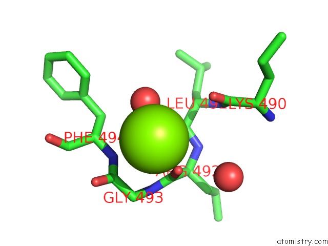
Mono view
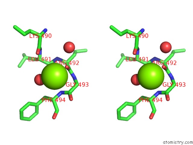
Stereo pair view
Mono view
Stereo pair view
A full contact list of Magnesium with other atoms in the Mg binding
site number 1 of Structure of Apo-Cuta within 5.0Å range:
|
Reference:
D.Malik,
K.Kobylecki,
P.Krawczyk,
J.Poznanski,
A.Jakielaszek,
A.Napiorkowska,
A.Dziembowski,
R.Tomecki,
M.Nowotny.
Structure and Mechanism of Cuta, Rna Nucleotidyl Transferase with An Unusual Preference For Cytosine. Nucleic Acids Res. V. 48 9387 2020.
ISSN: ESSN 1362-4962
PubMed: 32785623
DOI: 10.1093/NAR/GKAA647
Page generated: Wed Oct 2 01:12:14 2024
ISSN: ESSN 1362-4962
PubMed: 32785623
DOI: 10.1093/NAR/GKAA647
Last articles
Zn in 9J0NZn in 9J0O
Zn in 9J0P
Zn in 9FJX
Zn in 9EKB
Zn in 9C0F
Zn in 9CAH
Zn in 9CH0
Zn in 9CH3
Zn in 9CH1