Magnesium »
PDB 6ztc-7a1g »
6zvd »
Magnesium in PDB 6zvd: 14-3-3 Sigma in Complex with Phosphorylated GAB2PT391 Peptide - 96H Incubation
Protein crystallography data
The structure of 14-3-3 Sigma in Complex with Phosphorylated GAB2PT391 Peptide - 96H Incubation, PDB code: 6zvd
was solved by
A.Ballone,
R.A.Lau,
F.P.A.Zweipfenning,
C.Ottmann,
with X-Ray Crystallography technique. A brief refinement statistics is given in the table below:
| Resolution Low / High (Å) | 41.17 / 2.50 |
| Space group | C 2 2 21 |
| Cell size a, b, c (Å), α, β, γ (°) | 82.35, 111.984, 62.427, 90, 90, 90 |
| R / Rfree (%) | 17.4 / 23.4 |
Magnesium Binding Sites:
The binding sites of Magnesium atom in the 14-3-3 Sigma in Complex with Phosphorylated GAB2PT391 Peptide - 96H Incubation
(pdb code 6zvd). This binding sites where shown within
5.0 Angstroms radius around Magnesium atom.
In total 2 binding sites of Magnesium where determined in the 14-3-3 Sigma in Complex with Phosphorylated GAB2PT391 Peptide - 96H Incubation, PDB code: 6zvd:
Jump to Magnesium binding site number: 1; 2;
In total 2 binding sites of Magnesium where determined in the 14-3-3 Sigma in Complex with Phosphorylated GAB2PT391 Peptide - 96H Incubation, PDB code: 6zvd:
Jump to Magnesium binding site number: 1; 2;
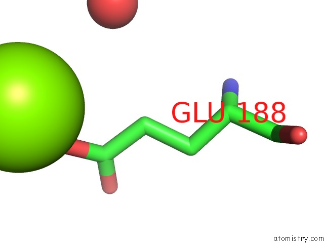
Magnesium binding site 1 out of 2 in 6zvd
Go back to
Magnesium binding site 1 out
of 2 in the 14-3-3 Sigma in Complex with Phosphorylated GAB2PT391 Peptide - 96H Incubation
Mono view

Stereo pair view
Mono view
Stereo pair view
A full contact list of Magnesium with other atoms in the Mg binding
site number 1 of 14-3-3 Sigma in Complex with Phosphorylated GAB2PT391 Peptide - 96H Incubation within 5.0Å range:
|
Magnesium binding site 2 out of 2 in 6zvd
Go back to
Magnesium binding site 2 out
of 2 in the 14-3-3 Sigma in Complex with Phosphorylated GAB2PT391 Peptide - 96H Incubation

Mono view

Stereo pair view
Mono view
Stereo pair view
A full contact list of Magnesium with other atoms in the Mg binding
site number 2 of 14-3-3 Sigma in Complex with Phosphorylated GAB2PT391 Peptide - 96H Incubation within 5.0Å range:
|
Reference:
A.Ballone,
R.A.Lau,
F.P.A.Zweipfenning,
C.Ottmann.
A New Soaking Procedure For X-Ray Crystallography Structural Determination of Protein-Peptide Complexes To Be Published.
Page generated: Wed Oct 2 03:12:32 2024
Last articles
Zn in 9JYWZn in 9IR4
Zn in 9IR3
Zn in 9GMX
Zn in 9GMW
Zn in 9JEJ
Zn in 9ERF
Zn in 9ERE
Zn in 9EGV
Zn in 9EGW