Magnesium »
PDB 6zup-7a1x »
7a0v »
Magnesium in PDB 7a0v: Crystal Structure of the 5-Phosphatase Domain of SYNAPTOJANIN1 in Complex with A Nanobody
Enzymatic activity of Crystal Structure of the 5-Phosphatase Domain of SYNAPTOJANIN1 in Complex with A Nanobody
All present enzymatic activity of Crystal Structure of the 5-Phosphatase Domain of SYNAPTOJANIN1 in Complex with A Nanobody:
3.1.3.36;
3.1.3.36;
Protein crystallography data
The structure of Crystal Structure of the 5-Phosphatase Domain of SYNAPTOJANIN1 in Complex with A Nanobody, PDB code: 7a0v
was solved by
J.Paesmans,
C.Galicia,
E.Martin,
W.Versees,
with X-Ray Crystallography technique. A brief refinement statistics is given in the table below:
| Resolution Low / High (Å) | 86.81 / 2.30 |
| Space group | C 1 2 1 |
| Cell size a, b, c (Å), α, β, γ (°) | 168.874, 108.793, 100.974, 90, 120.72, 90 |
| R / Rfree (%) | 19.6 / 25.2 |
Magnesium Binding Sites:
The binding sites of Magnesium atom in the Crystal Structure of the 5-Phosphatase Domain of SYNAPTOJANIN1 in Complex with A Nanobody
(pdb code 7a0v). This binding sites where shown within
5.0 Angstroms radius around Magnesium atom.
In total 3 binding sites of Magnesium where determined in the Crystal Structure of the 5-Phosphatase Domain of SYNAPTOJANIN1 in Complex with A Nanobody, PDB code: 7a0v:
Jump to Magnesium binding site number: 1; 2; 3;
In total 3 binding sites of Magnesium where determined in the Crystal Structure of the 5-Phosphatase Domain of SYNAPTOJANIN1 in Complex with A Nanobody, PDB code: 7a0v:
Jump to Magnesium binding site number: 1; 2; 3;
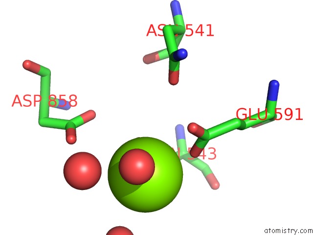
Magnesium binding site 1 out of 3 in 7a0v
Go back to
Magnesium binding site 1 out
of 3 in the Crystal Structure of the 5-Phosphatase Domain of SYNAPTOJANIN1 in Complex with A Nanobody

Mono view

Stereo pair view
Mono view
Stereo pair view
A full contact list of Magnesium with other atoms in the Mg binding
site number 1 of Crystal Structure of the 5-Phosphatase Domain of SYNAPTOJANIN1 in Complex with A Nanobody within 5.0Å range:
|
Magnesium binding site 2 out of 3 in 7a0v
Go back to
Magnesium binding site 2 out
of 3 in the Crystal Structure of the 5-Phosphatase Domain of SYNAPTOJANIN1 in Complex with A Nanobody
Mono view

Stereo pair view
Mono view
Stereo pair view
A full contact list of Magnesium with other atoms in the Mg binding
site number 2 of Crystal Structure of the 5-Phosphatase Domain of SYNAPTOJANIN1 in Complex with A Nanobody within 5.0Å range:
|
Magnesium binding site 3 out of 3 in 7a0v
Go back to
Magnesium binding site 3 out
of 3 in the Crystal Structure of the 5-Phosphatase Domain of SYNAPTOJANIN1 in Complex with A Nanobody

Mono view

Stereo pair view
Mono view
Stereo pair view
A full contact list of Magnesium with other atoms in the Mg binding
site number 3 of Crystal Structure of the 5-Phosphatase Domain of SYNAPTOJANIN1 in Complex with A Nanobody within 5.0Å range:
|
Reference:
J.Paesmans,
E.Martin,
B.Deckers,
M.Berghmans,
R.Sethi,
Y.Loeys,
E.Pardon,
J.Steyaert,
P.Verstreken,
C.Galicia,
W.Versees.
A Structure of Substrate-Bound SYNAPTOJANIN1 Provides New Insights in Its Mechanism and the Effect of Disease Mutations. Elife V. 9 2020.
ISSN: ESSN 2050-084X
PubMed: 33349335
DOI: 10.7554/ELIFE.64922
Page generated: Wed Oct 2 04:19:48 2024
ISSN: ESSN 2050-084X
PubMed: 33349335
DOI: 10.7554/ELIFE.64922
Last articles
Cl in 7TSPCl in 7TSO
Cl in 7TVG
Cl in 7TUY
Cl in 7TSN
Cl in 7TUO
Cl in 7TSM
Cl in 7TSL
Cl in 7TSK
Cl in 7TRV