Magnesium »
PDB 7bph-7c7a »
7c2b »
Magnesium in PDB 7c2b: Crystal Structure of Ferredoxin: Thioredoxin Reductase and Thioredoxin F2 Complex
Enzymatic activity of Crystal Structure of Ferredoxin: Thioredoxin Reductase and Thioredoxin F2 Complex
All present enzymatic activity of Crystal Structure of Ferredoxin: Thioredoxin Reductase and Thioredoxin F2 Complex:
1.8.7.2;
1.8.7.2;
Protein crystallography data
The structure of Crystal Structure of Ferredoxin: Thioredoxin Reductase and Thioredoxin F2 Complex, PDB code: 7c2b
was solved by
G.Kurisu,
L.Juniar,
H.Tanaka,
with X-Ray Crystallography technique. A brief refinement statistics is given in the table below:
| Resolution Low / High (Å) | 48.73 / 1.79 |
| Space group | P 21 21 21 |
| Cell size a, b, c (Å), α, β, γ (°) | 68.738, 83.943, 69.041, 90.00, 90.00, 90.00 |
| R / Rfree (%) | 17.9 / 21.1 |
Other elements in 7c2b:
The structure of Crystal Structure of Ferredoxin: Thioredoxin Reductase and Thioredoxin F2 Complex also contains other interesting chemical elements:
| Iron | (Fe) | 4 atoms |
Magnesium Binding Sites:
The binding sites of Magnesium atom in the Crystal Structure of Ferredoxin: Thioredoxin Reductase and Thioredoxin F2 Complex
(pdb code 7c2b). This binding sites where shown within
5.0 Angstroms radius around Magnesium atom.
In total only one binding site of Magnesium was determined in the Crystal Structure of Ferredoxin: Thioredoxin Reductase and Thioredoxin F2 Complex, PDB code: 7c2b:
In total only one binding site of Magnesium was determined in the Crystal Structure of Ferredoxin: Thioredoxin Reductase and Thioredoxin F2 Complex, PDB code: 7c2b:
Magnesium binding site 1 out of 1 in 7c2b
Go back to
Magnesium binding site 1 out
of 1 in the Crystal Structure of Ferredoxin: Thioredoxin Reductase and Thioredoxin F2 Complex
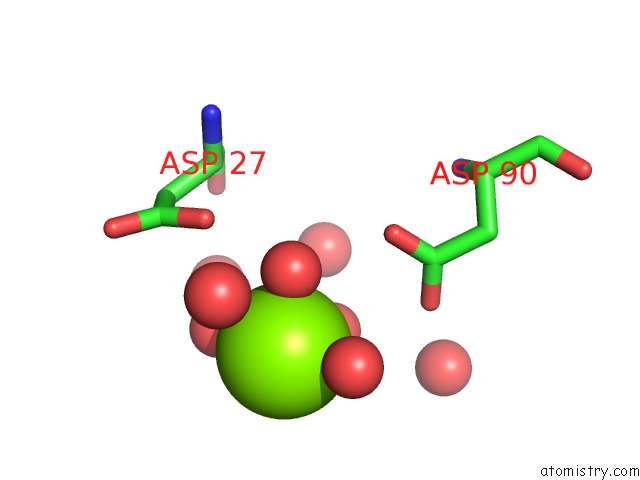
Mono view
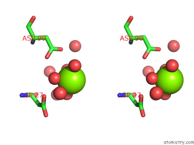
Stereo pair view
Mono view
Stereo pair view
A full contact list of Magnesium with other atoms in the Mg binding
site number 1 of Crystal Structure of Ferredoxin: Thioredoxin Reductase and Thioredoxin F2 Complex within 5.0Å range:
|
Reference:
L.Juniar,
H.Tanaka,
K.Yoshida,
T.Hisabori,
G.Kurisu.
Structural Basis For Thioredoxin Isoform-Based Fine-Tuning of Ferredoxin-Thioredoxin Reductase Activity. Protein Sci. V. 29 2538 2020.
ISSN: ESSN 1469-896X
PubMed: 33015914
DOI: 10.1002/PRO.3964
Page generated: Wed Oct 2 13:40:15 2024
ISSN: ESSN 1469-896X
PubMed: 33015914
DOI: 10.1002/PRO.3964
Last articles
Zn in 9J0NZn in 9J0O
Zn in 9J0P
Zn in 9FJX
Zn in 9EKB
Zn in 9C0F
Zn in 9CAH
Zn in 9CH0
Zn in 9CH3
Zn in 9CH1