Magnesium »
PDB 7c7j-7cji »
7cj5 »
Magnesium in PDB 7cj5: Crystal Structure of Homo Dimeric D-Allulose 3-Epimerase From Methylomonas Sp. in Complex with D-Fructose
Protein crystallography data
The structure of Crystal Structure of Homo Dimeric D-Allulose 3-Epimerase From Methylomonas Sp. in Complex with D-Fructose, PDB code: 7cj5
was solved by
H.Yoshida,
A.Yoshihara,
S.Kamitori,
with X-Ray Crystallography technique. A brief refinement statistics is given in the table below:
| Resolution Low / High (Å) | 19.94 / 1.80 |
| Space group | P 21 21 21 |
| Cell size a, b, c (Å), α, β, γ (°) | 45.42, 70.54, 140.22, 90, 90, 90 |
| R / Rfree (%) | 12.6 / 15.9 |
Other elements in 7cj5:
The structure of Crystal Structure of Homo Dimeric D-Allulose 3-Epimerase From Methylomonas Sp. in Complex with D-Fructose also contains other interesting chemical elements:
| Manganese | (Mn) | 2 atoms |
Magnesium Binding Sites:
The binding sites of Magnesium atom in the Crystal Structure of Homo Dimeric D-Allulose 3-Epimerase From Methylomonas Sp. in Complex with D-Fructose
(pdb code 7cj5). This binding sites where shown within
5.0 Angstroms radius around Magnesium atom.
In total 2 binding sites of Magnesium where determined in the Crystal Structure of Homo Dimeric D-Allulose 3-Epimerase From Methylomonas Sp. in Complex with D-Fructose, PDB code: 7cj5:
Jump to Magnesium binding site number: 1; 2;
In total 2 binding sites of Magnesium where determined in the Crystal Structure of Homo Dimeric D-Allulose 3-Epimerase From Methylomonas Sp. in Complex with D-Fructose, PDB code: 7cj5:
Jump to Magnesium binding site number: 1; 2;
Magnesium binding site 1 out of 2 in 7cj5
Go back to
Magnesium binding site 1 out
of 2 in the Crystal Structure of Homo Dimeric D-Allulose 3-Epimerase From Methylomonas Sp. in Complex with D-Fructose
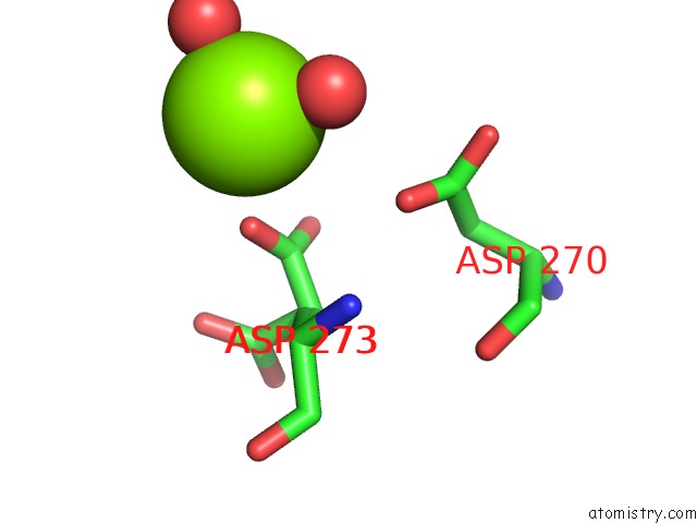
Mono view
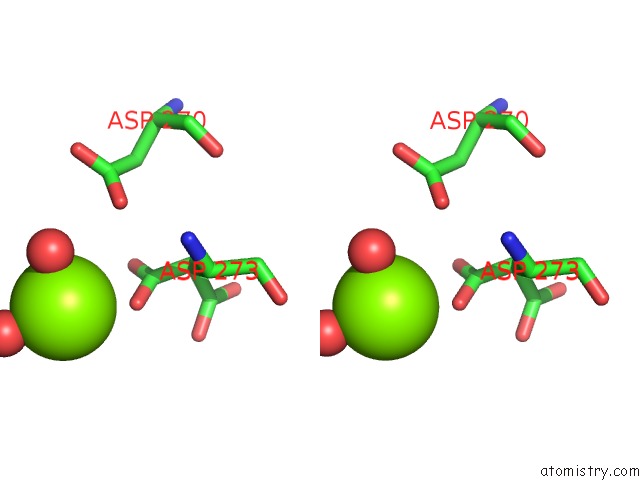
Stereo pair view
Mono view
Stereo pair view
A full contact list of Magnesium with other atoms in the Mg binding
site number 1 of Crystal Structure of Homo Dimeric D-Allulose 3-Epimerase From Methylomonas Sp. in Complex with D-Fructose within 5.0Å range:
|
Magnesium binding site 2 out of 2 in 7cj5
Go back to
Magnesium binding site 2 out
of 2 in the Crystal Structure of Homo Dimeric D-Allulose 3-Epimerase From Methylomonas Sp. in Complex with D-Fructose
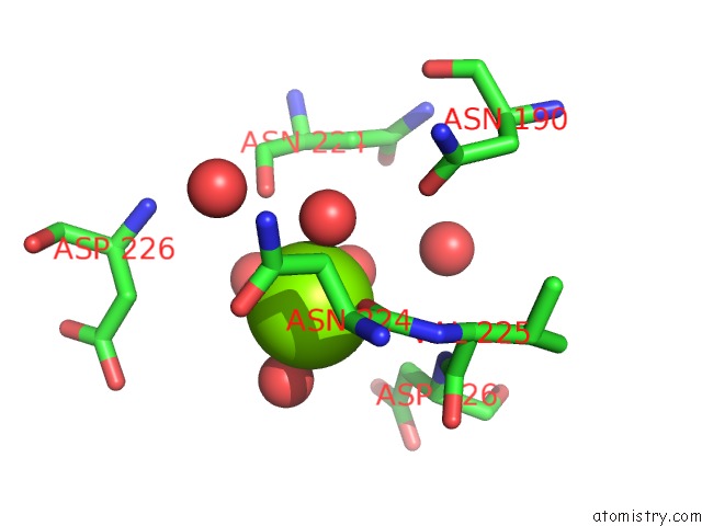
Mono view
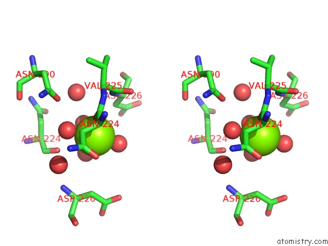
Stereo pair view
Mono view
Stereo pair view
A full contact list of Magnesium with other atoms in the Mg binding
site number 2 of Crystal Structure of Homo Dimeric D-Allulose 3-Epimerase From Methylomonas Sp. in Complex with D-Fructose within 5.0Å range:
|
Reference:
H.Yoshida,
A.Yoshihara,
S.Kato,
S.Mochizuki,
K.Akimitsu,
K.Izumori,
S.Kamitori.
Crystal Structure of A Novel Homodimeric L-Ribulose 3-Epimerase From Methylomonus Sp. Febs Open Bio 2021.
ISSN: ESSN 2211-5463
PubMed: 33838083
DOI: 10.1002/2211-5463.13159
Page generated: Wed Oct 2 14:04:32 2024
ISSN: ESSN 2211-5463
PubMed: 33838083
DOI: 10.1002/2211-5463.13159
Last articles
Zn in 9MJ5Zn in 9HNW
Zn in 9G0L
Zn in 9FNE
Zn in 9DZN
Zn in 9E0I
Zn in 9D32
Zn in 9DAK
Zn in 8ZXC
Zn in 8ZUF