Magnesium »
PDB 7diy-7dui »
7dld »
Magnesium in PDB 7dld: Crystal Structures of (S)-Carbonyl Reductases From Candida Parapsilosis in Different Oligomerization States
Protein crystallography data
The structure of Crystal Structures of (S)-Carbonyl Reductases From Candida Parapsilosis in Different Oligomerization States, PDB code: 7dld
was solved by
Y.H.Li,
R.Z.Zhang,
F.Forouhar,
C.Wang,
G.T.Montelione,
T.Szyperski,
Y.Xu,
J.F.Hunt,
with X-Ray Crystallography technique. A brief refinement statistics is given in the table below:
| Resolution Low / High (Å) | 43.22 / 1.75 |
| Space group | C 2 2 21 |
| Cell size a, b, c (Å), α, β, γ (°) | 69.262, 114.38, 126.371, 90, 90, 90 |
| R / Rfree (%) | 18.8 / 22.9 |
Magnesium Binding Sites:
The binding sites of Magnesium atom in the Crystal Structures of (S)-Carbonyl Reductases From Candida Parapsilosis in Different Oligomerization States
(pdb code 7dld). This binding sites where shown within
5.0 Angstroms radius around Magnesium atom.
In total 2 binding sites of Magnesium where determined in the Crystal Structures of (S)-Carbonyl Reductases From Candida Parapsilosis in Different Oligomerization States, PDB code: 7dld:
Jump to Magnesium binding site number: 1; 2;
In total 2 binding sites of Magnesium where determined in the Crystal Structures of (S)-Carbonyl Reductases From Candida Parapsilosis in Different Oligomerization States, PDB code: 7dld:
Jump to Magnesium binding site number: 1; 2;
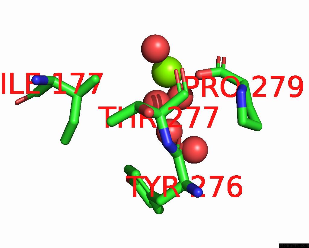
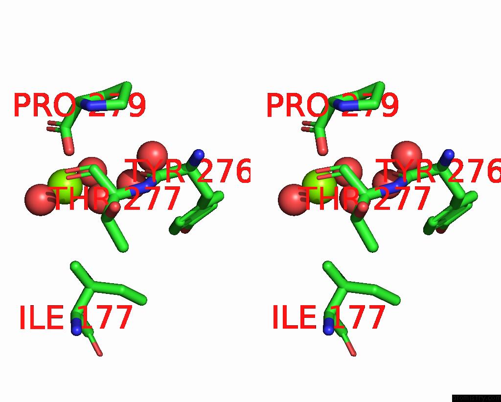
Magnesium binding site 1 out of 2 in 7dld
Go back to
Magnesium binding site 1 out
of 2 in the Crystal Structures of (S)-Carbonyl Reductases From Candida Parapsilosis in Different Oligomerization States

Mono view

Stereo pair view

Mono view

Stereo pair view
A full contact list of Magnesium with other atoms in the Mg binding
site number 1 of Crystal Structures of (S)-Carbonyl Reductases From Candida Parapsilosis in Different Oligomerization States within 5.0Å range:
|
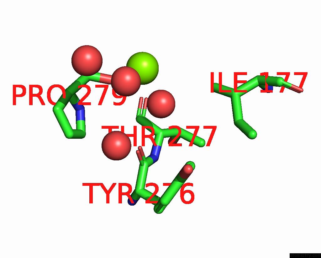
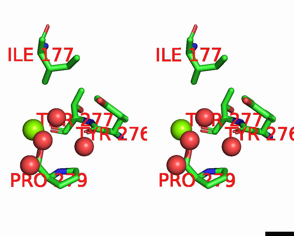
Magnesium binding site 2 out of 2 in 7dld
Go back to
Magnesium binding site 2 out
of 2 in the Crystal Structures of (S)-Carbonyl Reductases From Candida Parapsilosis in Different Oligomerization States

Mono view

Stereo pair view

Mono view

Stereo pair view
A full contact list of Magnesium with other atoms in the Mg binding
site number 2 of Crystal Structures of (S)-Carbonyl Reductases From Candida Parapsilosis in Different Oligomerization States within 5.0Å range:
|
Reference:
Y.Li,
R.Zhang,
C.Wang,
F.Forouhar,
O.B.Clarke,
S.Vorobiev,
S.Singh,
G.T.Montelione,
T.Szyperski,
Y.Xu,
J.F.Hunt.
Oligomeric Interactions Maintain Active-Site Structure in A Noncooperative Enzyme Family. Embo J. V. 41 08368 2022.
ISSN: ESSN 1460-2075
PubMed: 35801308
DOI: 10.15252/EMBJ.2021108368
Page generated: Wed Oct 2 15:55:00 2024
ISSN: ESSN 1460-2075
PubMed: 35801308
DOI: 10.15252/EMBJ.2021108368
Last articles
F in 7KHLF in 7KHH
F in 7KHG
F in 7KH6
F in 7KBG
F in 7KBN
F in 7KBC
F in 7KAF
F in 7KA1
F in 7K77