Magnesium »
PDB 7dic-7du3 »
7dqv »
Magnesium in PDB 7dqv: Crystal Structure of A CMABCB1 Mutant
Protein crystallography data
The structure of Crystal Structure of A CMABCB1 Mutant, PDB code: 7dqv
was solved by
K.Matsuoka,
T.Nakatsu,
H.Kato,
with X-Ray Crystallography technique. A brief refinement statistics is given in the table below:
| Resolution Low / High (Å) | 48.70 / 2.15 |
| Space group | P 41 3 2 |
| Cell size a, b, c (Å), α, β, γ (°) | 175.667, 175.667, 175.667, 90, 90, 90 |
| R / Rfree (%) | 18.5 / 21 |
Magnesium Binding Sites:
The binding sites of Magnesium atom in the Crystal Structure of A CMABCB1 Mutant
(pdb code 7dqv). This binding sites where shown within
5.0 Angstroms radius around Magnesium atom.
In total only one binding site of Magnesium was determined in the Crystal Structure of A CMABCB1 Mutant, PDB code: 7dqv:
In total only one binding site of Magnesium was determined in the Crystal Structure of A CMABCB1 Mutant, PDB code: 7dqv:
Magnesium binding site 1 out of 1 in 7dqv
Go back to
Magnesium binding site 1 out
of 1 in the Crystal Structure of A CMABCB1 Mutant
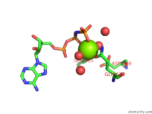
Mono view
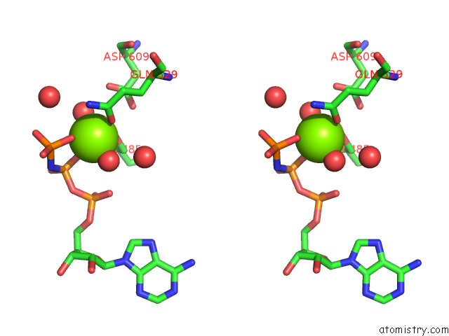
Stereo pair view
Mono view
Stereo pair view
A full contact list of Magnesium with other atoms in the Mg binding
site number 1 of Crystal Structure of A CMABCB1 Mutant within 5.0Å range:
|
Reference:
K.Matsuoka,
T.Nakatsu,
H.Kato.
The Crystal Structure of the CMABCB1 G132V Mutant, Which Favors the Outward-Facing State, Reveals the Mechanism of the Pivotal Joint Between TM1 and TM3. Protein Sci. 2021.
ISSN: ESSN 1469-896X
PubMed: 33683740
DOI: 10.1002/PRO.4058
Page generated: Wed Oct 2 15:59:12 2024
ISSN: ESSN 1469-896X
PubMed: 33683740
DOI: 10.1002/PRO.4058
Last articles
Zn in 9JYWZn in 9IR4
Zn in 9IR3
Zn in 9GMX
Zn in 9GMW
Zn in 9JEJ
Zn in 9ERF
Zn in 9ERE
Zn in 9EGV
Zn in 9EGW