Magnesium »
PDB 7duj-7e20 »
7dwr »
Magnesium in PDB 7dwr: Structure of Sulfolobus Solfataricus Sega-Adp Complex Bound to Dna
Protein crystallography data
The structure of Structure of Sulfolobus Solfataricus Sega-Adp Complex Bound to Dna, PDB code: 7dwr
was solved by
C.Y.Yen,
M.G.Lin,
C.D.Hsiao,
Y.J.Sun,
with X-Ray Crystallography technique. A brief refinement statistics is given in the table below:
| Resolution Low / High (Å) | 27.68 / 2.80 |
| Space group | C 1 2 1 |
| Cell size a, b, c (Å), α, β, γ (°) | 220, 60.541, 132.143, 90, 127.01, 90 |
| R / Rfree (%) | 23.9 / 29.8 |
Magnesium Binding Sites:
The binding sites of Magnesium atom in the Structure of Sulfolobus Solfataricus Sega-Adp Complex Bound to Dna
(pdb code 7dwr). This binding sites where shown within
5.0 Angstroms radius around Magnesium atom.
In total 4 binding sites of Magnesium where determined in the Structure of Sulfolobus Solfataricus Sega-Adp Complex Bound to Dna, PDB code: 7dwr:
Jump to Magnesium binding site number: 1; 2; 3; 4;
In total 4 binding sites of Magnesium where determined in the Structure of Sulfolobus Solfataricus Sega-Adp Complex Bound to Dna, PDB code: 7dwr:
Jump to Magnesium binding site number: 1; 2; 3; 4;
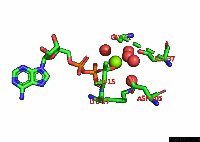
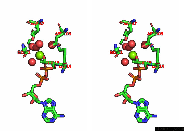
Magnesium binding site 1 out of 4 in 7dwr
Go back to
Magnesium binding site 1 out
of 4 in the Structure of Sulfolobus Solfataricus Sega-Adp Complex Bound to Dna
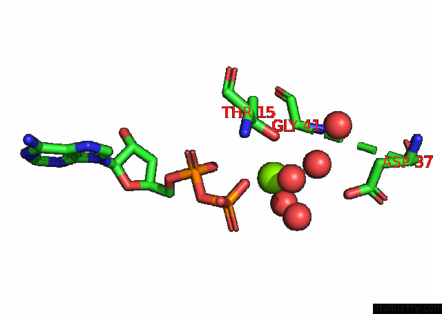
Mono view
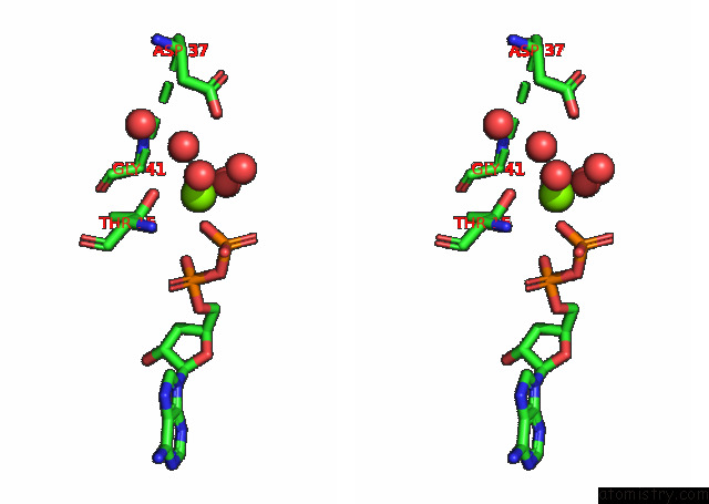
Stereo pair view
Mono view
Stereo pair view
A full contact list of Magnesium with other atoms in the Mg binding
site number 1 of Structure of Sulfolobus Solfataricus Sega-Adp Complex Bound to Dna within 5.0Å range:
|
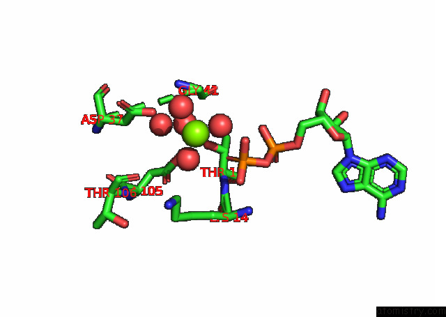
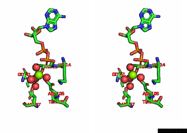
Magnesium binding site 2 out of 4 in 7dwr
Go back to
Magnesium binding site 2 out
of 4 in the Structure of Sulfolobus Solfataricus Sega-Adp Complex Bound to Dna
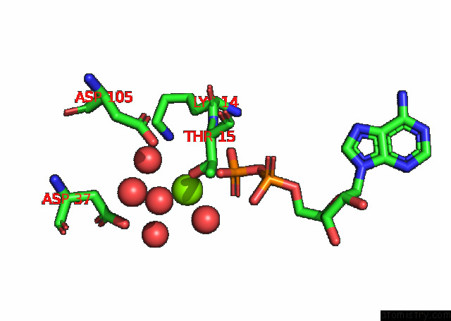
Mono view
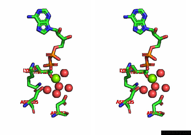
Stereo pair view
Mono view
Stereo pair view
A full contact list of Magnesium with other atoms in the Mg binding
site number 2 of Structure of Sulfolobus Solfataricus Sega-Adp Complex Bound to Dna within 5.0Å range:
|
Magnesium binding site 3 out of 4 in 7dwr
Go back to
Magnesium binding site 3 out
of 4 in the Structure of Sulfolobus Solfataricus Sega-Adp Complex Bound to Dna
Mono view
Stereo pair view
Mono view
Stereo pair view
A full contact list of Magnesium with other atoms in the Mg binding
site number 3 of Structure of Sulfolobus Solfataricus Sega-Adp Complex Bound to Dna within 5.0Å range:
|
Magnesium binding site 4 out of 4 in 7dwr
Go back to
Magnesium binding site 4 out
of 4 in the Structure of Sulfolobus Solfataricus Sega-Adp Complex Bound to Dna
Mono view
Stereo pair view
Mono view
Stereo pair view
A full contact list of Magnesium with other atoms in the Mg binding
site number 4 of Structure of Sulfolobus Solfataricus Sega-Adp Complex Bound to Dna within 5.0Å range:
|
Reference:
C.Y.Yen,
M.G.Lin,
B.W.Chen,
I.W.Ng,
N.Read,
A.F.Kabli,
C.T.Wu,
Y.Y.Shen,
C.H.Chen,
D.Barilla,
Y.J.Sun,
C.D.Hsiao.
Chromosome Segregation in Archaea: Sega- and Segb-Dna Complex Structures Provide Insights Into Segrosome Assembly. Nucleic Acids Res. 2021.
ISSN: ESSN 1362-4962
PubMed: 34850144
DOI: 10.1093/NAR/GKAB1155
Page generated: Wed Oct 2 16:32:29 2024
ISSN: ESSN 1362-4962
PubMed: 34850144
DOI: 10.1093/NAR/GKAB1155
Last articles
Zn in 9MJ5Zn in 9HNW
Zn in 9G0L
Zn in 9FNE
Zn in 9DZN
Zn in 9E0I
Zn in 9D32
Zn in 9DAK
Zn in 8ZXC
Zn in 8ZUF