Magnesium »
PDB 7e20-7eg0 »
7e6g »
Magnesium in PDB 7e6g: Crystal Structure of Diguanylate Cyclase Siad in Complex with Its Activator Siac From Pseudomonas Aeruginosa
Protein crystallography data
The structure of Crystal Structure of Diguanylate Cyclase Siad in Complex with Its Activator Siac From Pseudomonas Aeruginosa, PDB code: 7e6g
was solved by
J.S.Zhou,
L.Zhang,
L.Zhang,
with X-Ray Crystallography technique. A brief refinement statistics is given in the table below:
| Resolution Low / High (Å) | 46.30 / 2.65 |
| Space group | C 2 2 21 |
| Cell size a, b, c (Å), α, β, γ (°) | 82.525, 236.728, 148.627, 90, 90, 90 |
| R / Rfree (%) | 21.8 / 26.5 |
Magnesium Binding Sites:
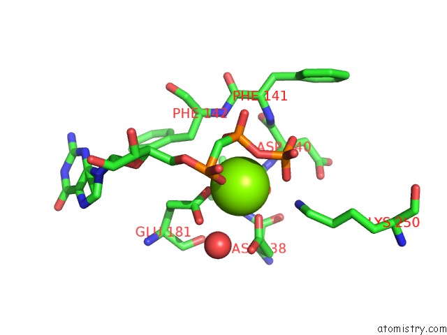
The binding sites of Magnesium atom in the Crystal Structure of Diguanylate Cyclase Siad in Complex with Its Activator Siac From Pseudomonas Aeruginosa
(pdb code 7e6g). This binding sites where shown within
5.0 Angstroms radius around Magnesium atom.
In total only one binding site of Magnesium was determined in the Crystal Structure of Diguanylate Cyclase Siad in Complex with Its Activator Siac From Pseudomonas Aeruginosa, PDB code: 7e6g:
In total only one binding site of Magnesium was determined in the Crystal Structure of Diguanylate Cyclase Siad in Complex with Its Activator Siac From Pseudomonas Aeruginosa, PDB code: 7e6g:
Magnesium binding site 1 out of 1 in 7e6g
Go back to
Magnesium binding site 1 out
of 1 in the Crystal Structure of Diguanylate Cyclase Siad in Complex with Its Activator Siac From Pseudomonas Aeruginosa
Mono view
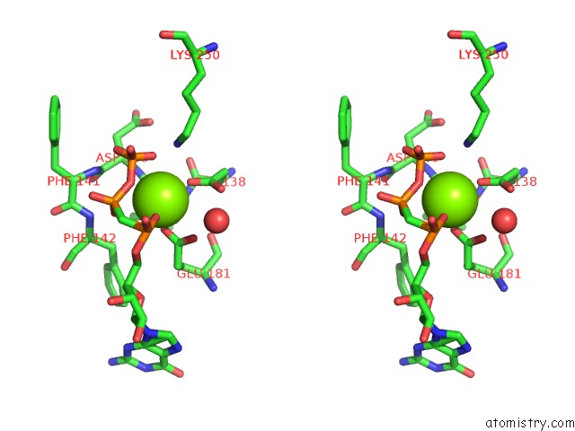
Stereo pair view
Mono view
Stereo pair view
A full contact list of Magnesium with other atoms in the Mg binding
site number 1 of Crystal Structure of Diguanylate Cyclase Siad in Complex with Its Activator Siac From Pseudomonas Aeruginosa within 5.0Å range:
|
Reference:
G.Chen,
J.Zhou,
Y.Zuo,
W.Huo,
J.Peng,
M.Li,
Y.Zhang,
T.Wang,
L.Zhang,
L.Zhang,
H.Liang.
Structural Basis For Diguanylate Cyclase Activation By Its Binding Partner in Pseudomonas Aeruginosa . Elife V. 10 2021.
ISSN: ESSN 2050-084X
PubMed: 34498587
DOI: 10.7554/ELIFE.67289
Page generated: Wed Oct 2 20:32:25 2024
ISSN: ESSN 2050-084X
PubMed: 34498587
DOI: 10.7554/ELIFE.67289
Last articles
Zn in 9J0NZn in 9J0O
Zn in 9J0P
Zn in 9FJX
Zn in 9EKB
Zn in 9C0F
Zn in 9CAH
Zn in 9CH0
Zn in 9CH3
Zn in 9CH1