Magnesium »
PDB 7eg4-7ens »
7eis »
Magnesium in PDB 7eis: Crystal Structure of Chondroitin Abc Lyase I in Complex with Chondroitin Disaccharide 0S
Enzymatic activity of Crystal Structure of Chondroitin Abc Lyase I in Complex with Chondroitin Disaccharide 0S
All present enzymatic activity of Crystal Structure of Chondroitin Abc Lyase I in Complex with Chondroitin Disaccharide 0S:
4.2.2.20;
4.2.2.20;
Protein crystallography data
The structure of Crystal Structure of Chondroitin Abc Lyase I in Complex with Chondroitin Disaccharide 0S, PDB code: 7eis
was solved by
M.Takashima,
A.Miyanaga,
T.Eguchi,
with X-Ray Crystallography technique. A brief refinement statistics is given in the table below:
| Resolution Low / High (Å) | 48.98 / 2.50 |
| Space group | P 21 21 21 |
| Cell size a, b, c (Å), α, β, γ (°) | 48.786, 94.29, 228.989, 90, 90, 90 |
| R / Rfree (%) | 20.4 / 27.8 |
Magnesium Binding Sites:
The binding sites of Magnesium atom in the Crystal Structure of Chondroitin Abc Lyase I in Complex with Chondroitin Disaccharide 0S
(pdb code 7eis). This binding sites where shown within
5.0 Angstroms radius around Magnesium atom.
In total only one binding site of Magnesium was determined in the Crystal Structure of Chondroitin Abc Lyase I in Complex with Chondroitin Disaccharide 0S, PDB code: 7eis:
In total only one binding site of Magnesium was determined in the Crystal Structure of Chondroitin Abc Lyase I in Complex with Chondroitin Disaccharide 0S, PDB code: 7eis:
Magnesium binding site 1 out of 1 in 7eis
Go back to
Magnesium binding site 1 out
of 1 in the Crystal Structure of Chondroitin Abc Lyase I in Complex with Chondroitin Disaccharide 0S
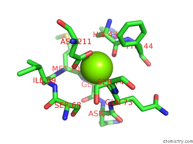
Mono view
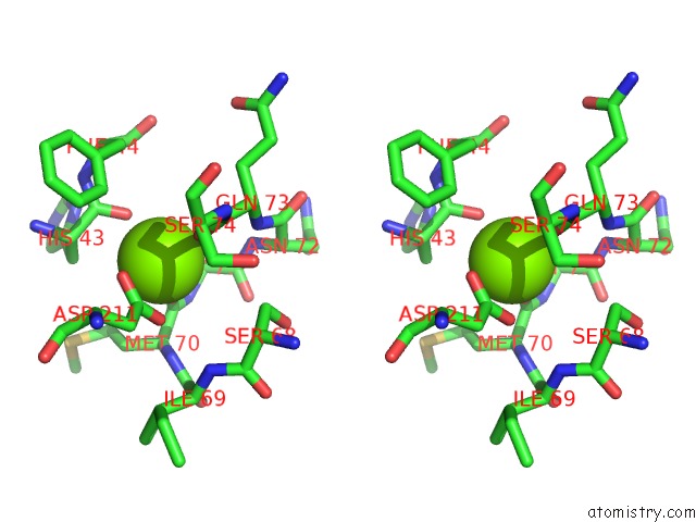
Stereo pair view
Mono view
Stereo pair view
A full contact list of Magnesium with other atoms in the Mg binding
site number 1 of Crystal Structure of Chondroitin Abc Lyase I in Complex with Chondroitin Disaccharide 0S within 5.0Å range:
|
Reference:
M.Takashima,
I.Watanabe,
A.Miyanaga,
T.Eguchi.
Substrate Specificity of Chondroitinase Abc I Based on Analyses of Biochemical Reactions and Crystal Structures in Complex with Disaccharides. Glycobiology 2021.
ISSN: ESSN 1460-2423
PubMed: 34392362
DOI: 10.1093/GLYCOB/CWAB086
Page generated: Wed Oct 2 20:44:34 2024
ISSN: ESSN 1460-2423
PubMed: 34392362
DOI: 10.1093/GLYCOB/CWAB086
Last articles
F in 7L7PF in 7L7O
F in 7L5E
F in 7L72
F in 7L5P
F in 7L69
F in 7L5O
F in 7L0K
F in 7L4W
F in 7L4U