Magnesium »
PDB 7ent-7evi »
7epq »
Magnesium in PDB 7epq: Crystal Structure of Exopolyphosphatase (Ppx) From Porphyromonas Gingivalis in Complex with Sulfate and Magnesium Ions
Protein crystallography data
The structure of Crystal Structure of Exopolyphosphatase (Ppx) From Porphyromonas Gingivalis in Complex with Sulfate and Magnesium Ions, PDB code: 7epq
was solved by
A.Zhang,
with X-Ray Crystallography technique. A brief refinement statistics is given in the table below:
| Resolution Low / High (Å) | 48.40 / 2.20 |
| Space group | P 21 21 21 |
| Cell size a, b, c (Å), α, β, γ (°) | 64.43, 86.61, 122.92, 90, 90, 90 |
| R / Rfree (%) | 18 / 24 |
Magnesium Binding Sites:
The binding sites of Magnesium atom in the Crystal Structure of Exopolyphosphatase (Ppx) From Porphyromonas Gingivalis in Complex with Sulfate and Magnesium Ions
(pdb code 7epq). This binding sites where shown within
5.0 Angstroms radius around Magnesium atom.
In total 7 binding sites of Magnesium where determined in the Crystal Structure of Exopolyphosphatase (Ppx) From Porphyromonas Gingivalis in Complex with Sulfate and Magnesium Ions, PDB code: 7epq:
Jump to Magnesium binding site number: 1; 2; 3; 4; 5; 6; 7;
In total 7 binding sites of Magnesium where determined in the Crystal Structure of Exopolyphosphatase (Ppx) From Porphyromonas Gingivalis in Complex with Sulfate and Magnesium Ions, PDB code: 7epq:
Jump to Magnesium binding site number: 1; 2; 3; 4; 5; 6; 7;
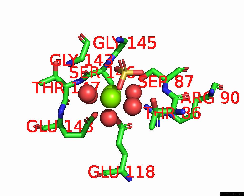
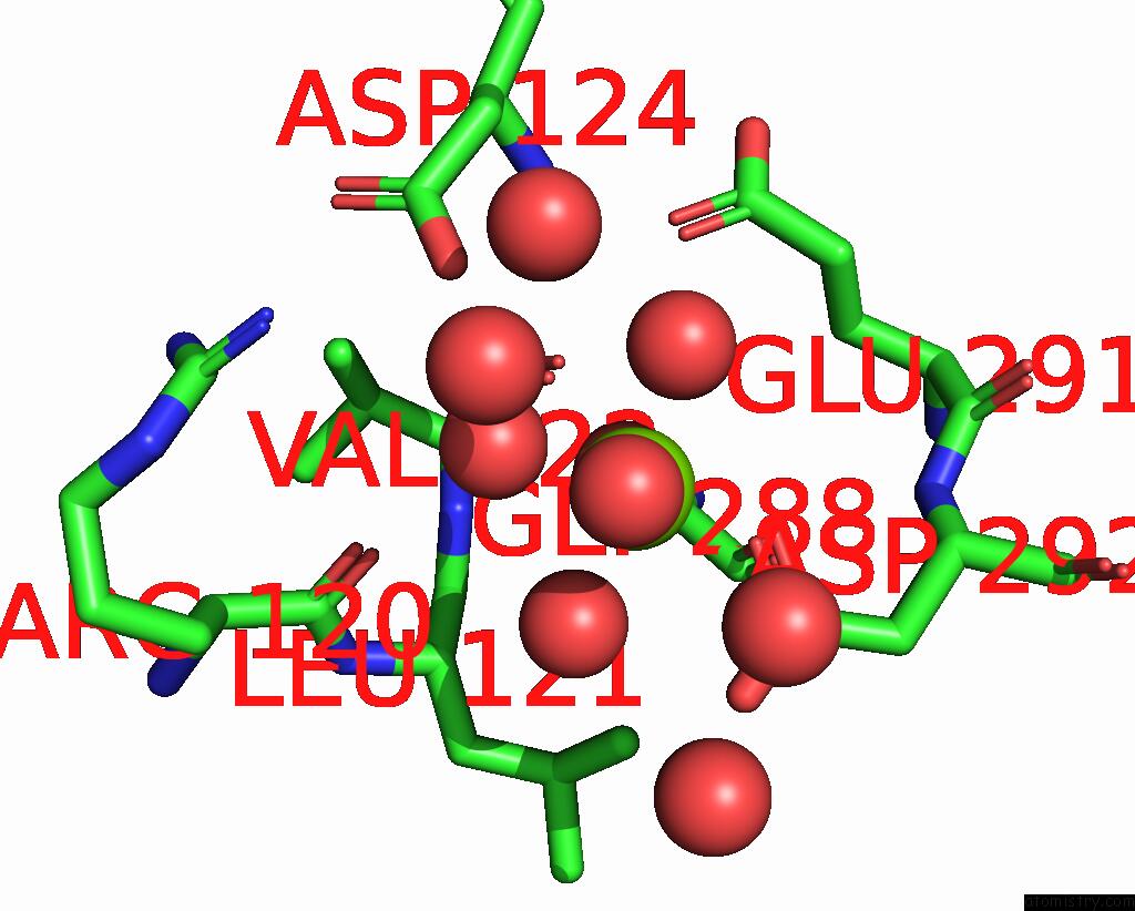
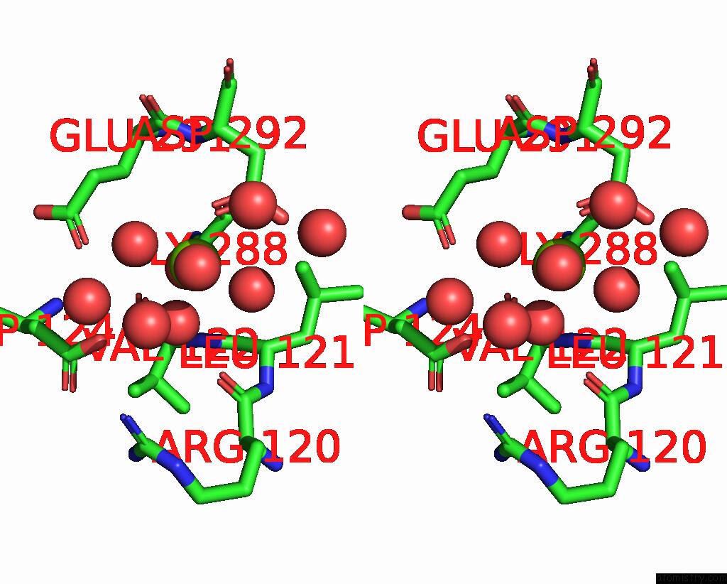
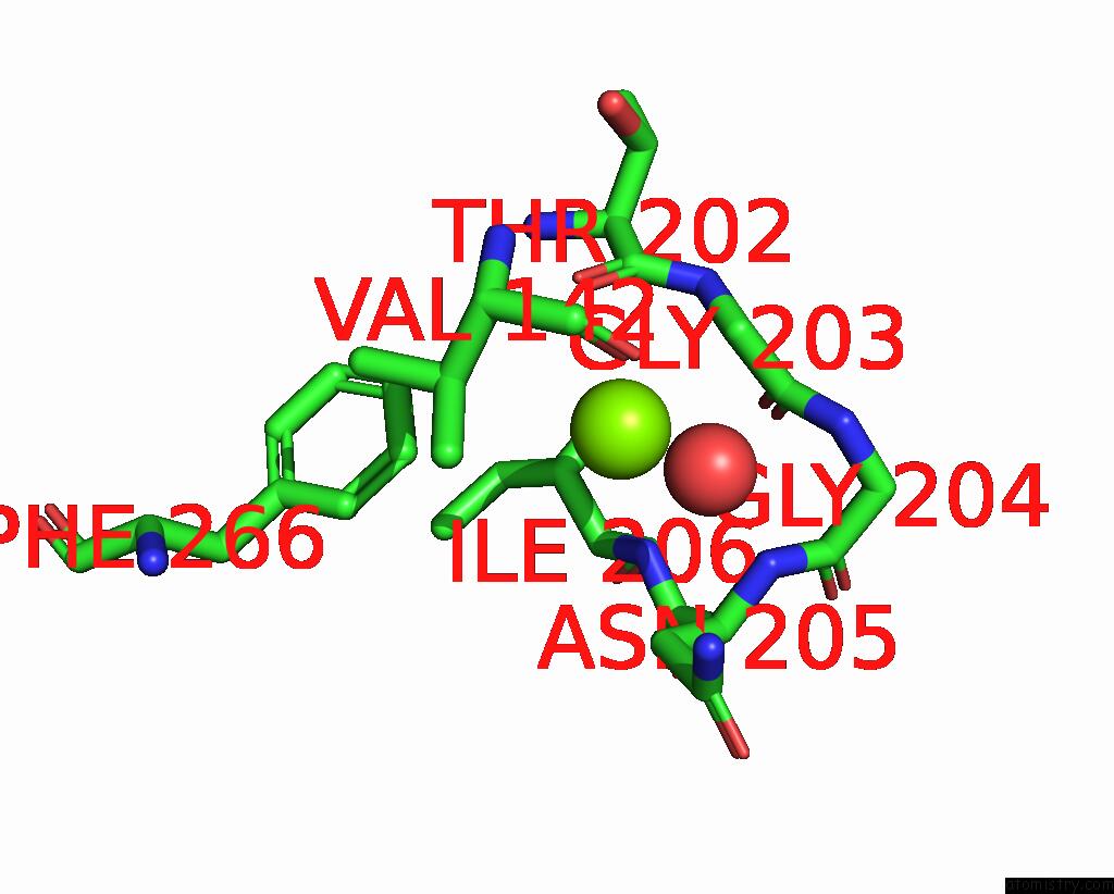
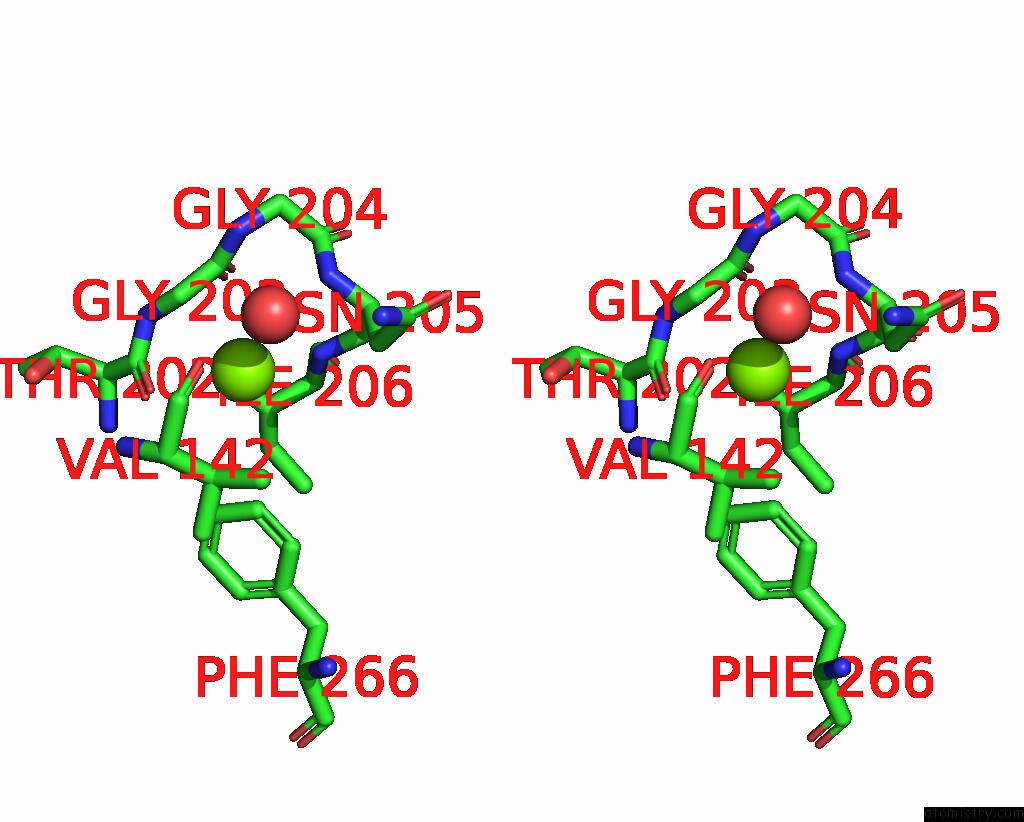
Magnesium binding site 1 out of 7 in 7epq
Go back to
Magnesium binding site 1 out
of 7 in the Crystal Structure of Exopolyphosphatase (Ppx) From Porphyromonas Gingivalis in Complex with Sulfate and Magnesium Ions
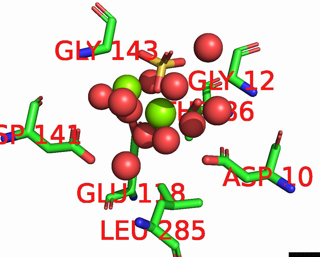
Mono view
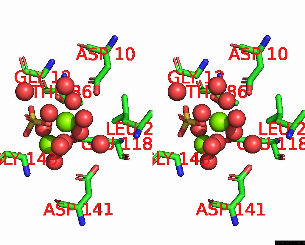
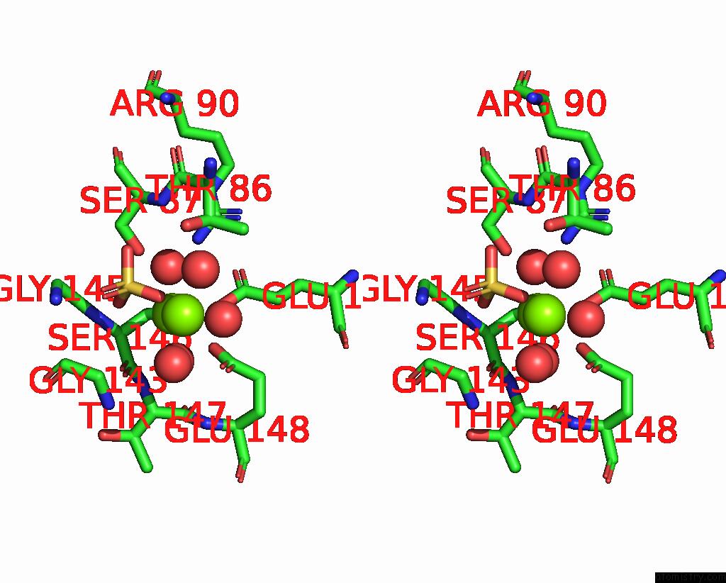
Stereo pair view
Mono view
Stereo pair view
A full contact list of Magnesium with other atoms in the Mg binding
site number 1 of Crystal Structure of Exopolyphosphatase (Ppx) From Porphyromonas Gingivalis in Complex with Sulfate and Magnesium Ions within 5.0Å range:
|
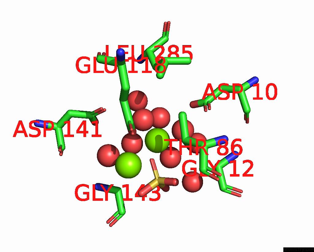
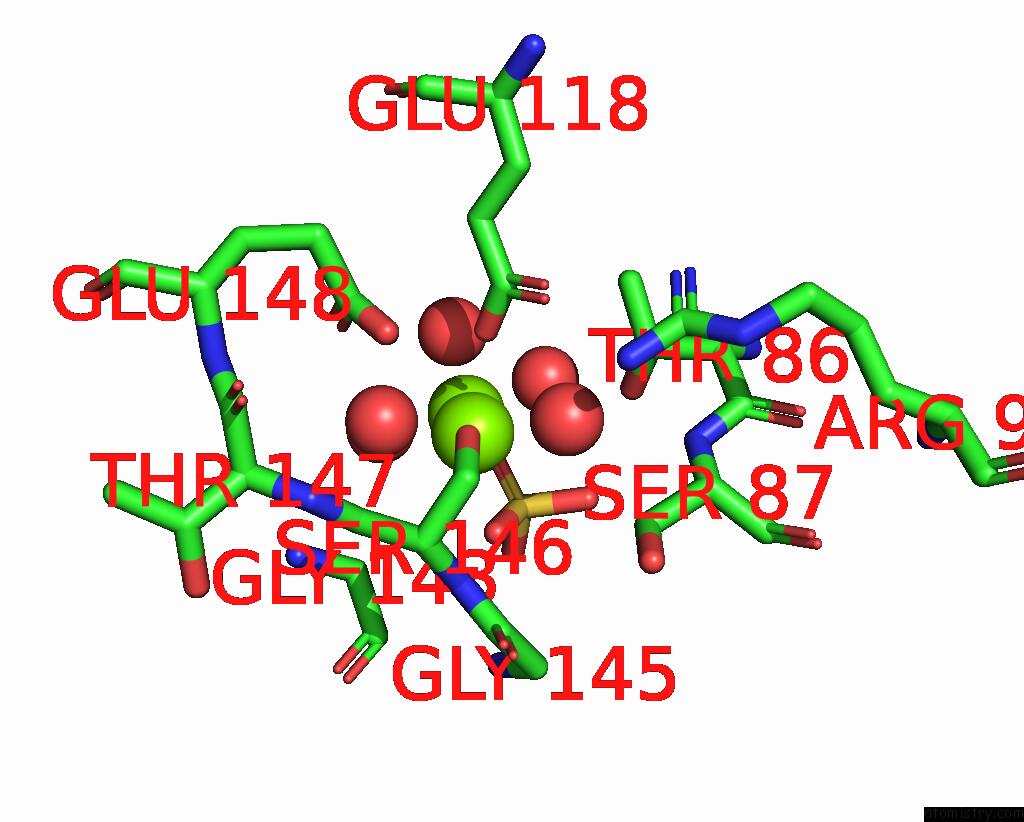
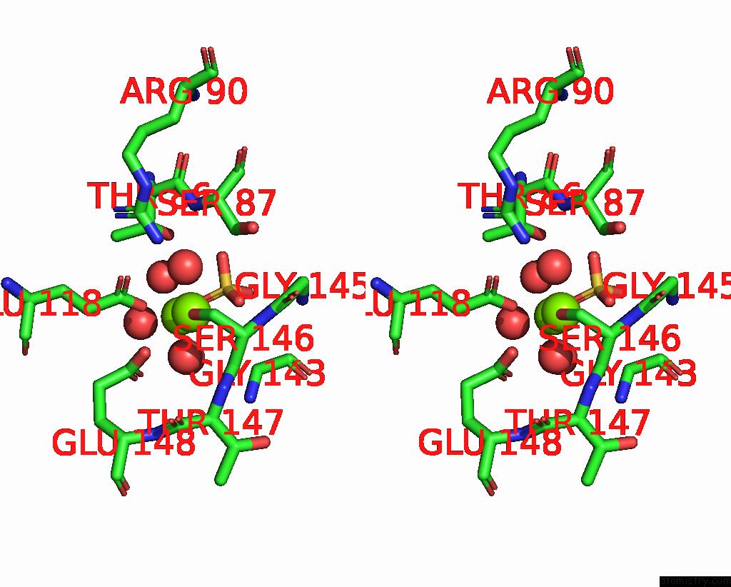
Magnesium binding site 2 out of 7 in 7epq
Go back to
Magnesium binding site 2 out
of 7 in the Crystal Structure of Exopolyphosphatase (Ppx) From Porphyromonas Gingivalis in Complex with Sulfate and Magnesium Ions
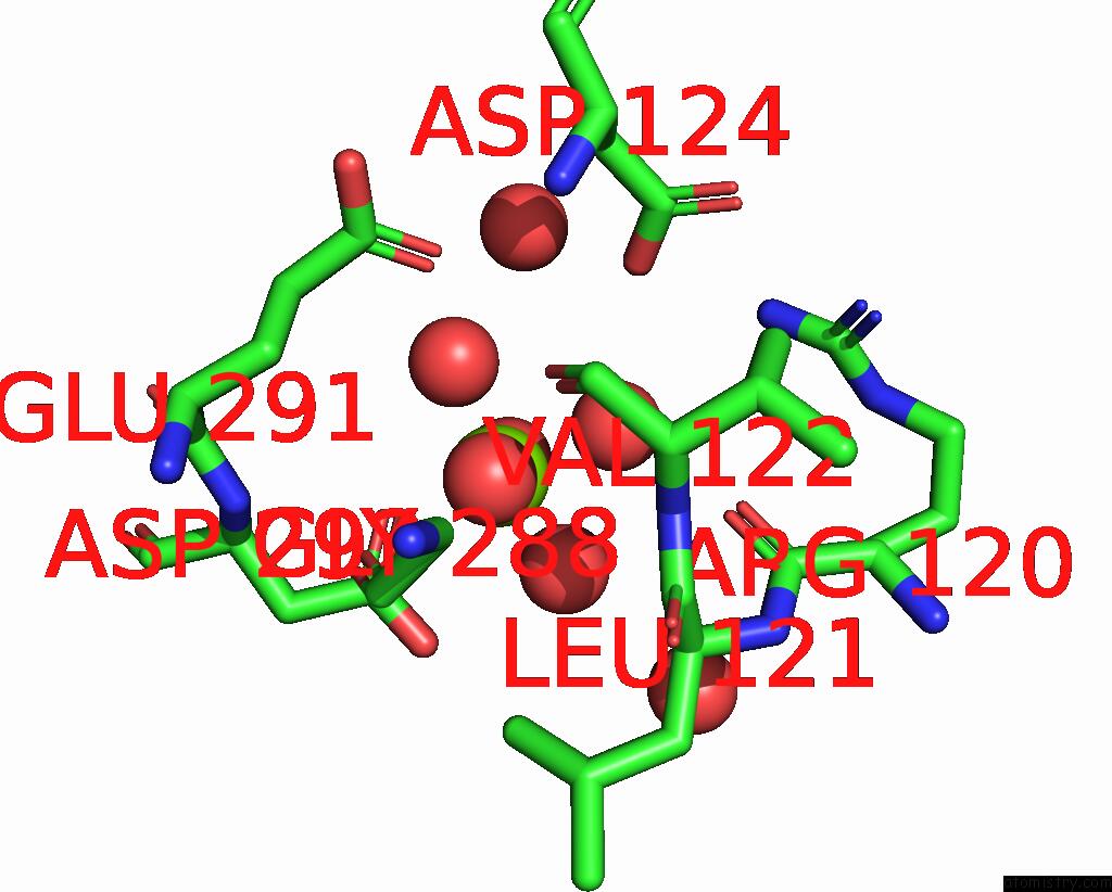
Mono view
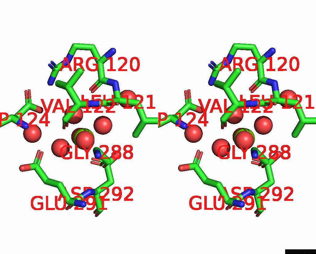
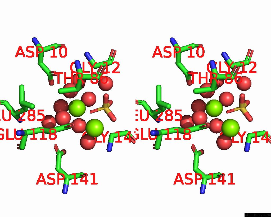
Stereo pair view
Mono view
Stereo pair view
A full contact list of Magnesium with other atoms in the Mg binding
site number 2 of Crystal Structure of Exopolyphosphatase (Ppx) From Porphyromonas Gingivalis in Complex with Sulfate and Magnesium Ions within 5.0Å range:
|
Magnesium binding site 3 out of 7 in 7epq
Go back to
Magnesium binding site 3 out
of 7 in the Crystal Structure of Exopolyphosphatase (Ppx) From Porphyromonas Gingivalis in Complex with Sulfate and Magnesium Ions
Mono view
Stereo pair view
Mono view
Stereo pair view
A full contact list of Magnesium with other atoms in the Mg binding
site number 3 of Crystal Structure of Exopolyphosphatase (Ppx) From Porphyromonas Gingivalis in Complex with Sulfate and Magnesium Ions within 5.0Å range:
|
Magnesium binding site 4 out of 7 in 7epq
Go back to
Magnesium binding site 4 out
of 7 in the Crystal Structure of Exopolyphosphatase (Ppx) From Porphyromonas Gingivalis in Complex with Sulfate and Magnesium Ions
Mono view
Stereo pair view
Mono view
Stereo pair view
A full contact list of Magnesium with other atoms in the Mg binding
site number 4 of Crystal Structure of Exopolyphosphatase (Ppx) From Porphyromonas Gingivalis in Complex with Sulfate and Magnesium Ions within 5.0Å range:
|
Magnesium binding site 5 out of 7 in 7epq
Go back to
Magnesium binding site 5 out
of 7 in the Crystal Structure of Exopolyphosphatase (Ppx) From Porphyromonas Gingivalis in Complex with Sulfate and Magnesium Ions
Mono view
Stereo pair view
Mono view
Stereo pair view
A full contact list of Magnesium with other atoms in the Mg binding
site number 5 of Crystal Structure of Exopolyphosphatase (Ppx) From Porphyromonas Gingivalis in Complex with Sulfate and Magnesium Ions within 5.0Å range:
|
Magnesium binding site 6 out of 7 in 7epq
Go back to
Magnesium binding site 6 out
of 7 in the Crystal Structure of Exopolyphosphatase (Ppx) From Porphyromonas Gingivalis in Complex with Sulfate and Magnesium Ions
Mono view
Stereo pair view
Mono view
Stereo pair view
A full contact list of Magnesium with other atoms in the Mg binding
site number 6 of Crystal Structure of Exopolyphosphatase (Ppx) From Porphyromonas Gingivalis in Complex with Sulfate and Magnesium Ions within 5.0Å range:
|
Magnesium binding site 7 out of 7 in 7epq
Go back to
Magnesium binding site 7 out
of 7 in the Crystal Structure of Exopolyphosphatase (Ppx) From Porphyromonas Gingivalis in Complex with Sulfate and Magnesium Ions
Mono view
Stereo pair view
Mono view
Stereo pair view
A full contact list of Magnesium with other atoms in the Mg binding
site number 7 of Crystal Structure of Exopolyphosphatase (Ppx) From Porphyromonas Gingivalis in Complex with Sulfate and Magnesium Ions within 5.0Å range:
|
Reference:
A.Zhang,
Z.Lu,
Y.Xu,
T.Qi,
W.Li,
L.Zhang,
Z.Cui.
The Structure of Exopolyphosphatase (Ppx) From Porphyromonas Gingivalis in Complex with Substrate Analogs and Magnesium Ions Reveals the Basis For Polyphosphate Processivity. J.Struct.Biol. V. 213 07767 2021.
ISSN: ESSN 1095-8657
PubMed: 34214602
DOI: 10.1016/J.JSB.2021.107767
Page generated: Wed Oct 2 20:54:56 2024
ISSN: ESSN 1095-8657
PubMed: 34214602
DOI: 10.1016/J.JSB.2021.107767
Last articles
Zn in 9MJ5Zn in 9HNW
Zn in 9G0L
Zn in 9FNE
Zn in 9DZN
Zn in 9E0I
Zn in 9D32
Zn in 9DAK
Zn in 8ZXC
Zn in 8ZUF