Magnesium »
PDB 7ent-7evi »
7eqj »
Magnesium in PDB 7eqj: Crystal Structure of E. Coli Valine Trna
Protein crystallography data
The structure of Crystal Structure of E. Coli Valine Trna, PDB code: 7eqj
was solved by
J.Kim,
H.Jeong,
with X-Ray Crystallography technique. A brief refinement statistics is given in the table below:
| Resolution Low / High (Å) | 29.49 / 2.04 |
| Space group | P 1 21 1 |
| Cell size a, b, c (Å), α, β, γ (°) | 53.541, 33.059, 131.797, 90, 98.15, 90 |
| R / Rfree (%) | 20.4 / 23.6 |
Other elements in 7eqj:
The structure of Crystal Structure of E. Coli Valine Trna also contains other interesting chemical elements:
| Sodium | (Na) | 4 atoms |
Magnesium Binding Sites:
The binding sites of Magnesium atom in the Crystal Structure of E. Coli Valine Trna
(pdb code 7eqj). This binding sites where shown within
5.0 Angstroms radius around Magnesium atom.
In total 6 binding sites of Magnesium where determined in the Crystal Structure of E. Coli Valine Trna, PDB code: 7eqj:
Jump to Magnesium binding site number: 1; 2; 3; 4; 5; 6;
In total 6 binding sites of Magnesium where determined in the Crystal Structure of E. Coli Valine Trna, PDB code: 7eqj:
Jump to Magnesium binding site number: 1; 2; 3; 4; 5; 6;
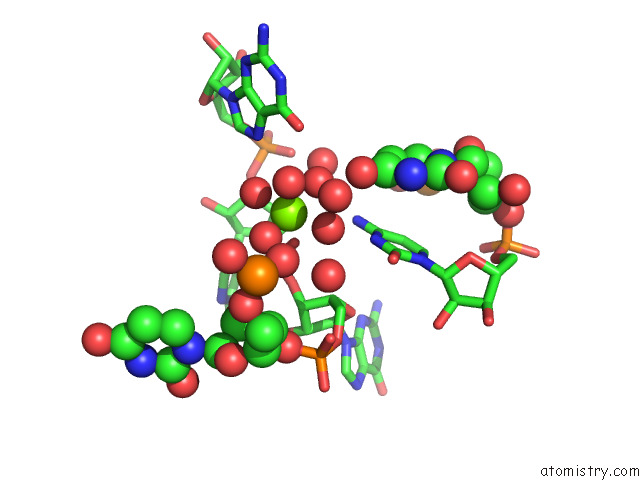
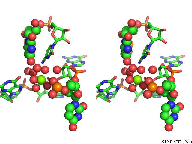
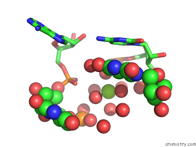
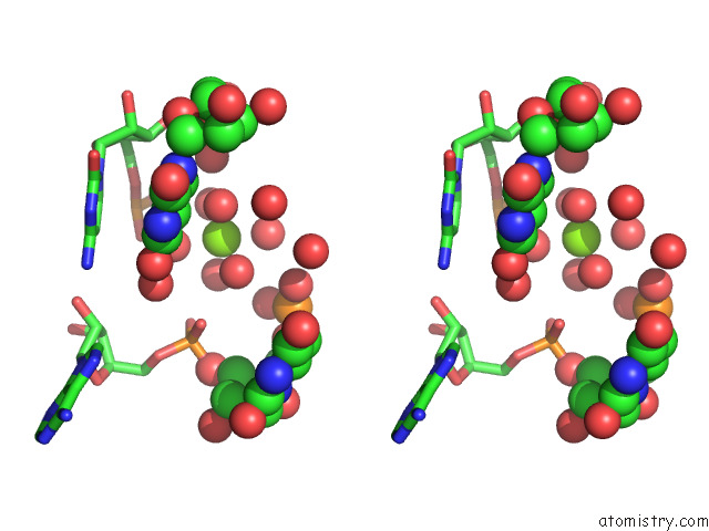
Magnesium binding site 1 out of 6 in 7eqj
Go back to
Magnesium binding site 1 out
of 6 in the Crystal Structure of E. Coli Valine Trna
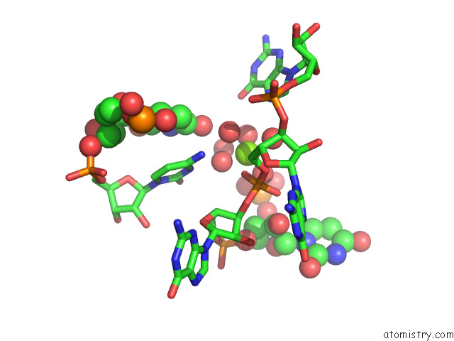
Mono view
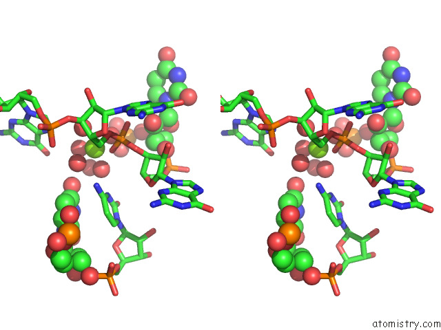
Stereo pair view
Mono view
Stereo pair view
A full contact list of Magnesium with other atoms in the Mg binding
site number 1 of Crystal Structure of E. Coli Valine Trna within 5.0Å range:
|
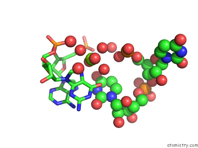
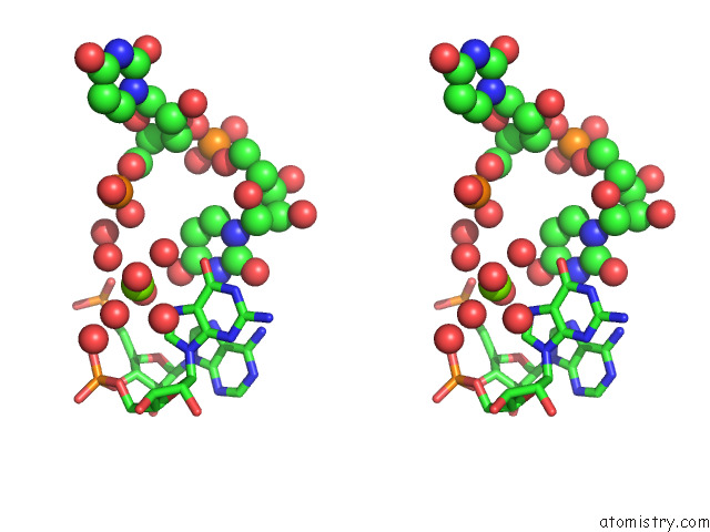
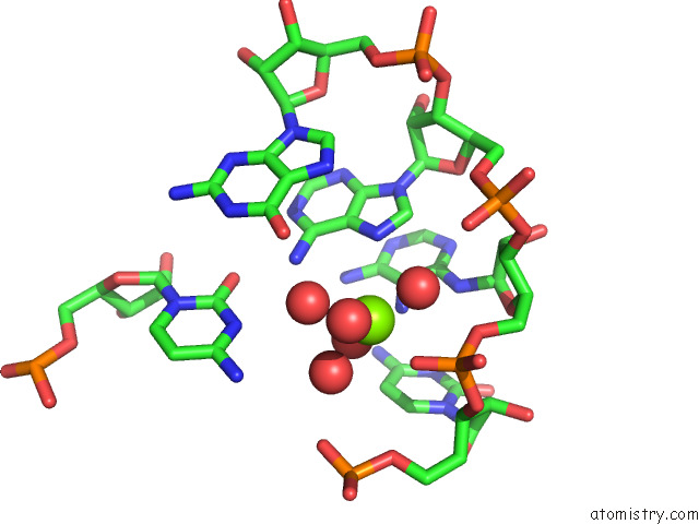
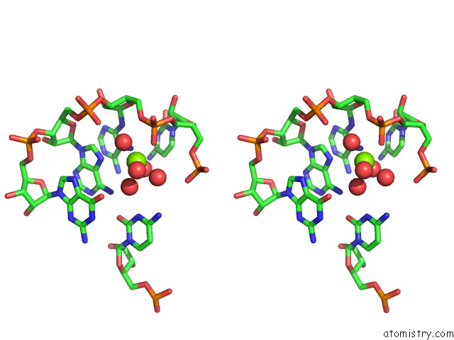
Magnesium binding site 2 out of 6 in 7eqj
Go back to
Magnesium binding site 2 out
of 6 in the Crystal Structure of E. Coli Valine Trna
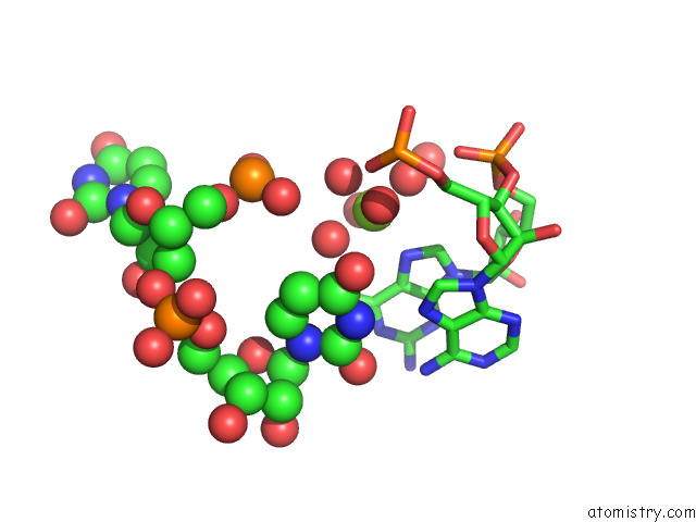
Mono view
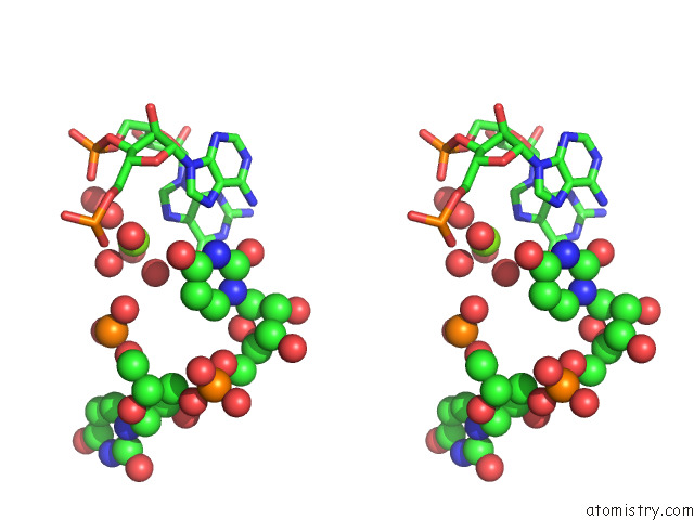
Stereo pair view
Mono view
Stereo pair view
A full contact list of Magnesium with other atoms in the Mg binding
site number 2 of Crystal Structure of E. Coli Valine Trna within 5.0Å range:
|
Magnesium binding site 3 out of 6 in 7eqj
Go back to
Magnesium binding site 3 out
of 6 in the Crystal Structure of E. Coli Valine Trna
Mono view
Stereo pair view
Mono view
Stereo pair view
A full contact list of Magnesium with other atoms in the Mg binding
site number 3 of Crystal Structure of E. Coli Valine Trna within 5.0Å range:
|
Magnesium binding site 4 out of 6 in 7eqj
Go back to
Magnesium binding site 4 out
of 6 in the Crystal Structure of E. Coli Valine Trna
Mono view
Stereo pair view
Mono view
Stereo pair view
A full contact list of Magnesium with other atoms in the Mg binding
site number 4 of Crystal Structure of E. Coli Valine Trna within 5.0Å range:
|
Magnesium binding site 5 out of 6 in 7eqj
Go back to
Magnesium binding site 5 out
of 6 in the Crystal Structure of E. Coli Valine Trna
Mono view
Stereo pair view
Mono view
Stereo pair view
A full contact list of Magnesium with other atoms in the Mg binding
site number 5 of Crystal Structure of E. Coli Valine Trna within 5.0Å range:
|
Magnesium binding site 6 out of 6 in 7eqj
Go back to
Magnesium binding site 6 out
of 6 in the Crystal Structure of E. Coli Valine Trna
Mono view
Stereo pair view
Mono view
Stereo pair view
A full contact list of Magnesium with other atoms in the Mg binding
site number 6 of Crystal Structure of E. Coli Valine Trna within 5.0Å range:
|
Reference:
H.Jeong,
J.Kim.
Unique Anticodon Loop Conformation with the Flipped-Out Wobble Nucleotide in the Crystal Structure of Unbound Trna Val . Rna V. 27 1330 2021.
ISSN: ESSN 1469-9001
PubMed: 34315814
DOI: 10.1261/RNA.078863.121
Page generated: Wed Oct 2 20:55:29 2024
ISSN: ESSN 1469-9001
PubMed: 34315814
DOI: 10.1261/RNA.078863.121
Last articles
Fe in 2YXOFe in 2YRS
Fe in 2YXC
Fe in 2YNM
Fe in 2YVJ
Fe in 2YP1
Fe in 2YU2
Fe in 2YU1
Fe in 2YQB
Fe in 2YOO