Magnesium »
PDB 7ew6-7f6j »
7f4x »
Magnesium in PDB 7f4x: Joint Neutron and X-Ray Crystal Structure of the Nucleotide-Binding Domain of HSP72 in Complex with Adp
Protein crystallography data
The structure of Joint Neutron and X-Ray Crystal Structure of the Nucleotide-Binding Domain of HSP72 in Complex with Adp, PDB code: 7f4x
was solved by
T.Yokoyama,
A.Ostermann,
T.E.Schrader,
with X-Ray Crystallography technique. A brief refinement statistics is given in the table below:
| Resolution Low / High (Å) | N/A / 1.60 |
| Space group | P 21 21 21 |
| Cell size a, b, c (Å), α, β, γ (°) | 46.677, 64.618, 145.593, 90, 90, 90 |
| R / Rfree (%) | 18.4 / 22.2 |
Other elements in 7f4x:
The structure of Joint Neutron and X-Ray Crystal Structure of the Nucleotide-Binding Domain of HSP72 in Complex with Adp also contains other interesting chemical elements:
| Sodium | (Na) | 1 atom |
Magnesium Binding Sites:
The binding sites of Magnesium atom in the Joint Neutron and X-Ray Crystal Structure of the Nucleotide-Binding Domain of HSP72 in Complex with Adp
(pdb code 7f4x). This binding sites where shown within
5.0 Angstroms radius around Magnesium atom.
In total only one binding site of Magnesium was determined in the Joint Neutron and X-Ray Crystal Structure of the Nucleotide-Binding Domain of HSP72 in Complex with Adp, PDB code: 7f4x:
In total only one binding site of Magnesium was determined in the Joint Neutron and X-Ray Crystal Structure of the Nucleotide-Binding Domain of HSP72 in Complex with Adp, PDB code: 7f4x:
Magnesium binding site 1 out of 1 in 7f4x
Go back to
Magnesium binding site 1 out
of 1 in the Joint Neutron and X-Ray Crystal Structure of the Nucleotide-Binding Domain of HSP72 in Complex with Adp
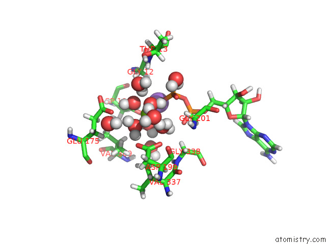
Mono view
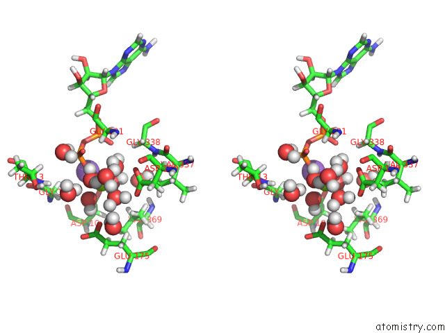
Stereo pair view
Mono view
Stereo pair view
A full contact list of Magnesium with other atoms in the Mg binding
site number 1 of Joint Neutron and X-Ray Crystal Structure of the Nucleotide-Binding Domain of HSP72 in Complex with Adp within 5.0Å range:
|
Reference:
T.Yokoyama,
S.Fujii,
A.Ostermann,
T.E.Schrader,
Y.Nabeshima,
M.Mizuguchi.
Neutron Crystallographic Analysis of the Nucleotide-Binding Domain of HSP72 in Complex with Adp. Iucrj V. 9 562 2022.
ISSN: ESSN 2052-2525
DOI: 10.1107/S2052252522006297
Page generated: Wed Oct 2 21:10:51 2024
ISSN: ESSN 2052-2525
DOI: 10.1107/S2052252522006297
Last articles
Zn in 9MJ5Zn in 9HNW
Zn in 9G0L
Zn in 9FNE
Zn in 9DZN
Zn in 9E0I
Zn in 9D32
Zn in 9DAK
Zn in 8ZXC
Zn in 8ZUF