Magnesium »
PDB 7jpp-7k06 »
7jsn »
Magnesium in PDB 7jsn: Structure of the Visual Signaling Complex Between Transducin and Phosphodiesterase 6
Enzymatic activity of Structure of the Visual Signaling Complex Between Transducin and Phosphodiesterase 6
All present enzymatic activity of Structure of the Visual Signaling Complex Between Transducin and Phosphodiesterase 6:
3.1.4.35;
3.1.4.35;
Other elements in 7jsn:
The structure of Structure of the Visual Signaling Complex Between Transducin and Phosphodiesterase 6 also contains other interesting chemical elements:
| Zinc | (Zn) | 2 atoms |
Magnesium Binding Sites:
The binding sites of Magnesium atom in the Structure of the Visual Signaling Complex Between Transducin and Phosphodiesterase 6
(pdb code 7jsn). This binding sites where shown within
5.0 Angstroms radius around Magnesium atom.
In total 2 binding sites of Magnesium where determined in the Structure of the Visual Signaling Complex Between Transducin and Phosphodiesterase 6, PDB code: 7jsn:
Jump to Magnesium binding site number: 1; 2;
In total 2 binding sites of Magnesium where determined in the Structure of the Visual Signaling Complex Between Transducin and Phosphodiesterase 6, PDB code: 7jsn:
Jump to Magnesium binding site number: 1; 2;
Magnesium binding site 1 out of 2 in 7jsn
Go back to
Magnesium binding site 1 out
of 2 in the Structure of the Visual Signaling Complex Between Transducin and Phosphodiesterase 6
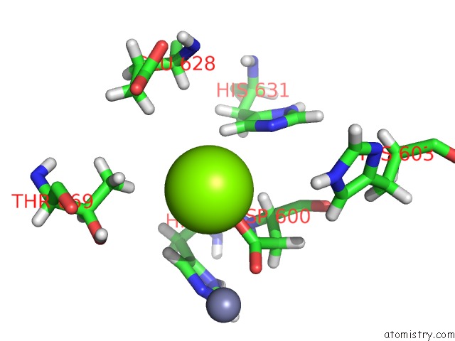
Mono view
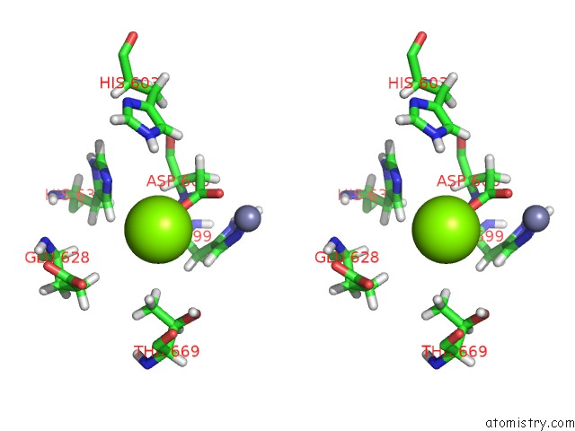
Stereo pair view
Mono view
Stereo pair view
A full contact list of Magnesium with other atoms in the Mg binding
site number 1 of Structure of the Visual Signaling Complex Between Transducin and Phosphodiesterase 6 within 5.0Å range:
|
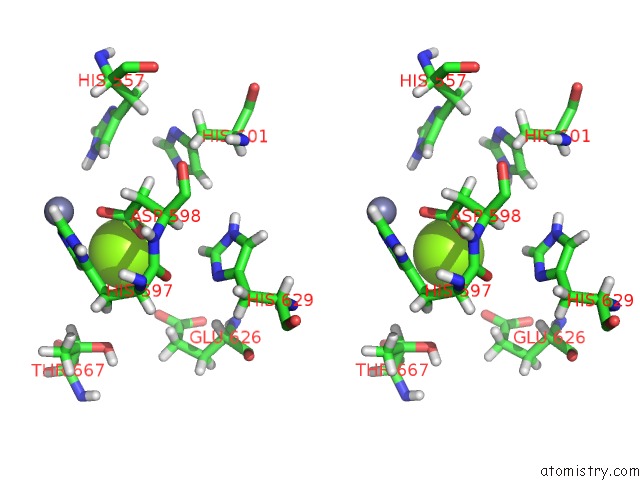
Magnesium binding site 2 out of 2 in 7jsn
Go back to
Magnesium binding site 2 out
of 2 in the Structure of the Visual Signaling Complex Between Transducin and Phosphodiesterase 6
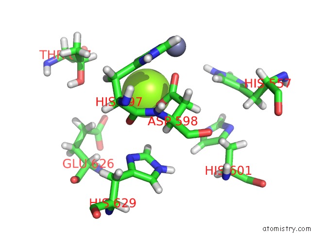
Mono view
Stereo pair view
Mono view
Stereo pair view
A full contact list of Magnesium with other atoms in the Mg binding
site number 2 of Structure of the Visual Signaling Complex Between Transducin and Phosphodiesterase 6 within 5.0Å range:
|
Reference:
Y.Gao,
G.Eskici,
S.Ramachandran,
F.Poitevin,
A.B.Seven,
O.Panova,
G.Skiniotis,
R.A.Cerione.
Structure of the Visual Signaling Complex Between Transducin and Phosphodiesterase 6. Mol.Cell V. 80 237 2020.
ISSN: ISSN 1097-2765
PubMed: 33007200
DOI: 10.1016/J.MOLCEL.2020.09.013
Page generated: Wed Oct 2 21:59:53 2024
ISSN: ISSN 1097-2765
PubMed: 33007200
DOI: 10.1016/J.MOLCEL.2020.09.013
Last articles
Zn in 9MJ5Zn in 9HNW
Zn in 9G0L
Zn in 9FNE
Zn in 9DZN
Zn in 9E0I
Zn in 9D32
Zn in 9DAK
Zn in 8ZXC
Zn in 8ZUF