Magnesium »
PDB 7klu-7ktf »
7koh »
Magnesium in PDB 7koh: Crystal Structure of Antigen 43 From Escherichia Coli EDL933
Protein crystallography data
The structure of Crystal Structure of Antigen 43 From Escherichia Coli EDL933, PDB code: 7koh
was solved by
J.L.Vo,
J.J.Paxman,
B.Heras,
with X-Ray Crystallography technique. A brief refinement statistics is given in the table below:
| Resolution Low / High (Å) | 48.78 / 2.98 |
| Space group | P 21 21 21 |
| Cell size a, b, c (Å), α, β, γ (°) | 97.55, 178.66, 246.23, 90, 90, 90 |
| R / Rfree (%) | 20.6 / 25.4 |
Other elements in 7koh:
The structure of Crystal Structure of Antigen 43 From Escherichia Coli EDL933 also contains other interesting chemical elements:
| Chlorine | (Cl) | 1 atom |
Magnesium Binding Sites:
The binding sites of Magnesium atom in the Crystal Structure of Antigen 43 From Escherichia Coli EDL933
(pdb code 7koh). This binding sites where shown within
5.0 Angstroms radius around Magnesium atom.
In total 3 binding sites of Magnesium where determined in the Crystal Structure of Antigen 43 From Escherichia Coli EDL933, PDB code: 7koh:
Jump to Magnesium binding site number: 1; 2; 3;
In total 3 binding sites of Magnesium where determined in the Crystal Structure of Antigen 43 From Escherichia Coli EDL933, PDB code: 7koh:
Jump to Magnesium binding site number: 1; 2; 3;
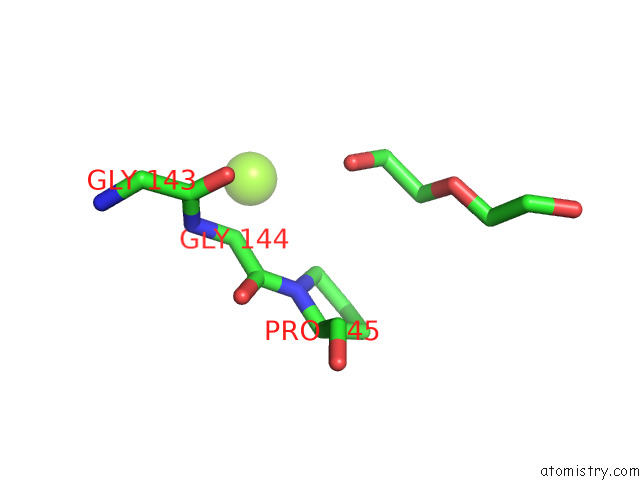
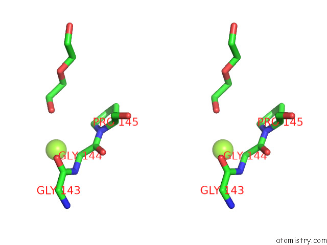
Magnesium binding site 1 out of 3 in 7koh
Go back to
Magnesium binding site 1 out
of 3 in the Crystal Structure of Antigen 43 From Escherichia Coli EDL933
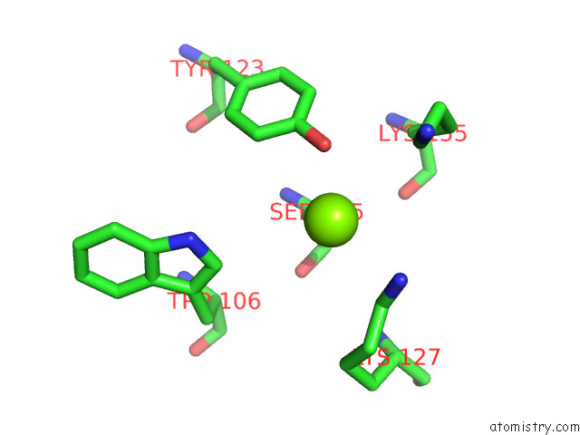
Mono view
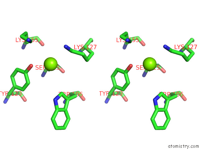
Stereo pair view
Mono view
Stereo pair view
A full contact list of Magnesium with other atoms in the Mg binding
site number 1 of Crystal Structure of Antigen 43 From Escherichia Coli EDL933 within 5.0Å range:
|
Magnesium binding site 2 out of 3 in 7koh
Go back to
Magnesium binding site 2 out
of 3 in the Crystal Structure of Antigen 43 From Escherichia Coli EDL933
Mono view
Stereo pair view
Mono view
Stereo pair view
A full contact list of Magnesium with other atoms in the Mg binding
site number 2 of Crystal Structure of Antigen 43 From Escherichia Coli EDL933 within 5.0Å range:
|
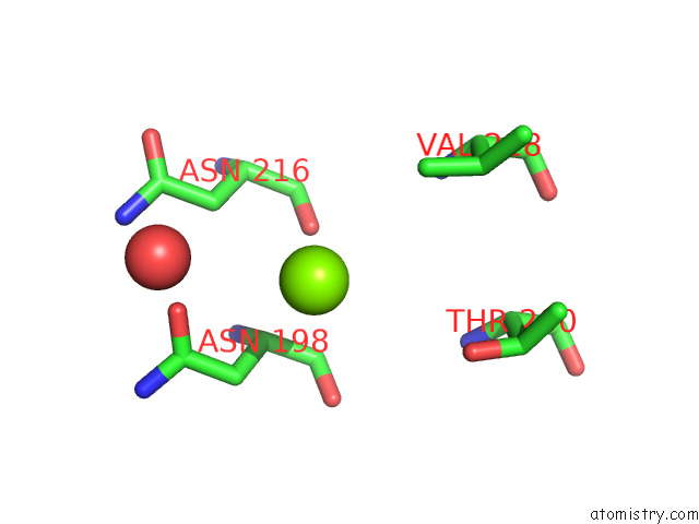
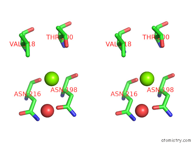
Magnesium binding site 3 out of 3 in 7koh
Go back to
Magnesium binding site 3 out
of 3 in the Crystal Structure of Antigen 43 From Escherichia Coli EDL933
Mono view
Stereo pair view
Mono view
Stereo pair view
A full contact list of Magnesium with other atoms in the Mg binding
site number 3 of Crystal Structure of Antigen 43 From Escherichia Coli EDL933 within 5.0Å range:
|
Reference:
J.L.Vo,
G.C.Martinez-Ortiz,
M.Totsika,
A.Lo,
A.E.Whitten,
L.Hor,
K.M.Peters,
V.Ageorges,
N.Caccia,
M.Desvaux,
M.A.Schembri,
J.J.Paxman,
B.B.Heras.
Fine Tuning the Mechanism of Antigen 43 Self-Association to Modulate Aggregation Levels of Escherichia Coli Pathogens To Be Published.
Page generated: Wed Oct 2 22:33:18 2024
Last articles
Ca in 5S5OCa in 5S5N
Ca in 5S5M
Ca in 5S5L
Ca in 5S5K
Ca in 5S5J
Ca in 5S5I
Ca in 5S5H
Ca in 5S5G
Ca in 5S5F