Magnesium »
PDB 7mo0-7mz8 »
7msr »
Magnesium in PDB 7msr: Human E105QA Gtp-Specific Succinyl-Coa Synthetase Complexed with Succinyl-Phosphate, Magnesium Ion and Desulfo-Coenzyme A
Enzymatic activity of Human E105QA Gtp-Specific Succinyl-Coa Synthetase Complexed with Succinyl-Phosphate, Magnesium Ion and Desulfo-Coenzyme A
All present enzymatic activity of Human E105QA Gtp-Specific Succinyl-Coa Synthetase Complexed with Succinyl-Phosphate, Magnesium Ion and Desulfo-Coenzyme A:
6.2.1.4; 6.2.1.5;
6.2.1.4; 6.2.1.5;
Protein crystallography data
The structure of Human E105QA Gtp-Specific Succinyl-Coa Synthetase Complexed with Succinyl-Phosphate, Magnesium Ion and Desulfo-Coenzyme A, PDB code: 7msr
was solved by
J.Huang,
M.E.Fraser,
with X-Ray Crystallography technique. A brief refinement statistics is given in the table below:
| Resolution Low / High (Å) | 39.13 / 1.58 |
| Space group | P 1 21 1 |
| Cell size a, b, c (Å), α, β, γ (°) | 80.436, 81.452, 54.331, 90, 103.37, 90 |
| R / Rfree (%) | 19.9 / 22.1 |
Magnesium Binding Sites:
The binding sites of Magnesium atom in the Human E105QA Gtp-Specific Succinyl-Coa Synthetase Complexed with Succinyl-Phosphate, Magnesium Ion and Desulfo-Coenzyme A
(pdb code 7msr). This binding sites where shown within
5.0 Angstroms radius around Magnesium atom.
In total only one binding site of Magnesium was determined in the Human E105QA Gtp-Specific Succinyl-Coa Synthetase Complexed with Succinyl-Phosphate, Magnesium Ion and Desulfo-Coenzyme A, PDB code: 7msr:
In total only one binding site of Magnesium was determined in the Human E105QA Gtp-Specific Succinyl-Coa Synthetase Complexed with Succinyl-Phosphate, Magnesium Ion and Desulfo-Coenzyme A, PDB code: 7msr:
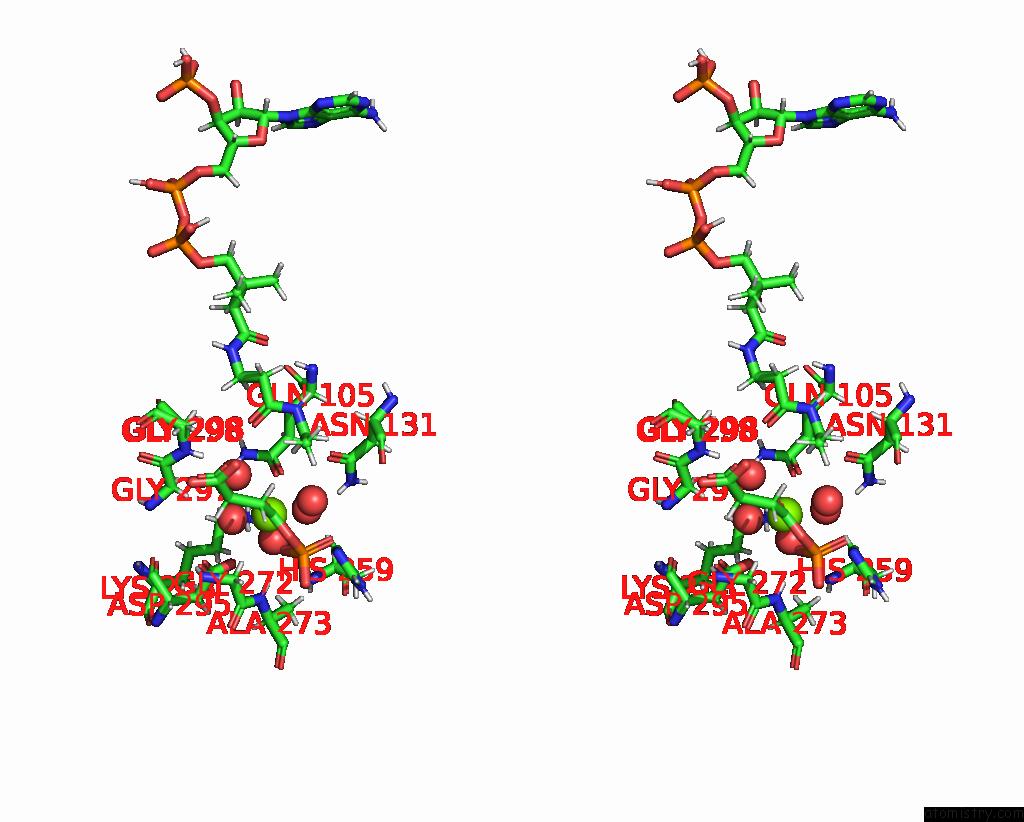
Magnesium binding site 1 out of 1 in 7msr
Go back to
Magnesium binding site 1 out
of 1 in the Human E105QA Gtp-Specific Succinyl-Coa Synthetase Complexed with Succinyl-Phosphate, Magnesium Ion and Desulfo-Coenzyme A

Mono view
Stereo pair view
Mono view
Stereo pair view
A full contact list of Magnesium with other atoms in the Mg binding
site number 1 of Human E105QA Gtp-Specific Succinyl-Coa Synthetase Complexed with Succinyl-Phosphate, Magnesium Ion and Desulfo-Coenzyme A within 5.0Å range:
|
Reference:
J.Huang,
M.E.Fraser.
The Structure of Succinyl-Coa Synthetase Bound to the Succinyl-Phosphate Intermediate Clarifies the Catalytic Mechanism of Atp-Citrate Lyase Acta Crystallogr.,Sect.F V. 78 363 2022.
ISSN: ESSN 2053-230X
DOI: 10.1107/S2053230X22008810
Page generated: Thu Oct 3 01:07:55 2024
ISSN: ESSN 2053-230X
DOI: 10.1107/S2053230X22008810
Last articles
F in 4GLUF in 4GNK
F in 4GOA
F in 4GM8
F in 4GMX
F in 4GLX
F in 4G3B
F in 4GL9
F in 4G4M
F in 4GJ3