Magnesium »
PDB 7mo0-7mz8 »
7mxb »
Magnesium in PDB 7mxb: Crystal Structure of the S/T Protein Kinase Pkng From Corynebacterium Glutamicum in Complex with Amp-Pnp
Protein crystallography data
The structure of Crystal Structure of the S/T Protein Kinase Pkng From Corynebacterium Glutamicum in Complex with Amp-Pnp, PDB code: 7mxb
was solved by
M.N.Lisa,
P.M.Alzari,
with X-Ray Crystallography technique. A brief refinement statistics is given in the table below:
| Resolution Low / High (Å) | 43.02 / 2.20 |
| Space group | P 1 21 1 |
| Cell size a, b, c (Å), α, β, γ (°) | 104.663, 42.741, 175.327, 90, 95.31, 90 |
| R / Rfree (%) | 20.9 / 24.6 |
Magnesium Binding Sites:
The binding sites of Magnesium atom in the Crystal Structure of the S/T Protein Kinase Pkng From Corynebacterium Glutamicum in Complex with Amp-Pnp
(pdb code 7mxb). This binding sites where shown within
5.0 Angstroms radius around Magnesium atom.
In total 6 binding sites of Magnesium where determined in the Crystal Structure of the S/T Protein Kinase Pkng From Corynebacterium Glutamicum in Complex with Amp-Pnp, PDB code: 7mxb:
Jump to Magnesium binding site number: 1; 2; 3; 4; 5; 6;
In total 6 binding sites of Magnesium where determined in the Crystal Structure of the S/T Protein Kinase Pkng From Corynebacterium Glutamicum in Complex with Amp-Pnp, PDB code: 7mxb:
Jump to Magnesium binding site number: 1; 2; 3; 4; 5; 6;
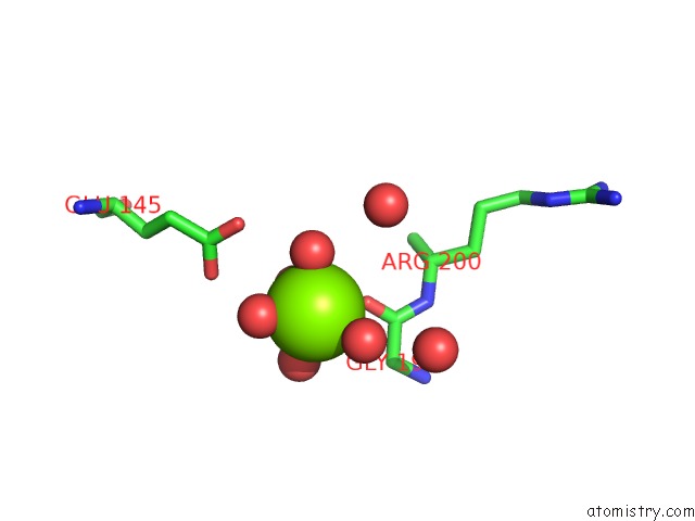
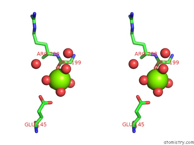
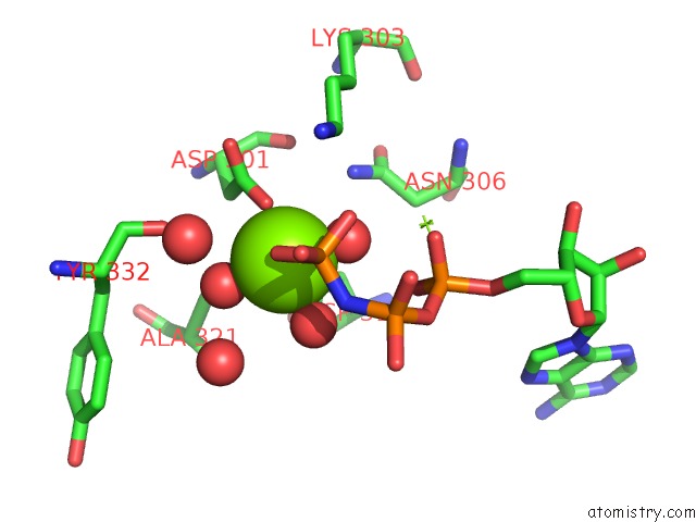
Magnesium binding site 1 out of 6 in 7mxb
Go back to
Magnesium binding site 1 out
of 6 in the Crystal Structure of the S/T Protein Kinase Pkng From Corynebacterium Glutamicum in Complex with Amp-Pnp
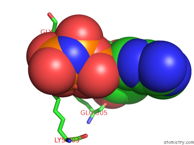
Mono view
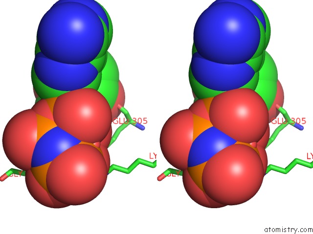
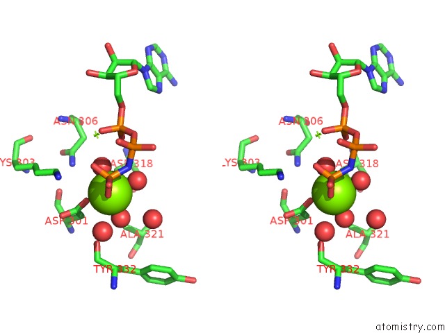
Stereo pair view
Mono view
Stereo pair view
A full contact list of Magnesium with other atoms in the Mg binding
site number 1 of Crystal Structure of the S/T Protein Kinase Pkng From Corynebacterium Glutamicum in Complex with Amp-Pnp within 5.0Å range:
|
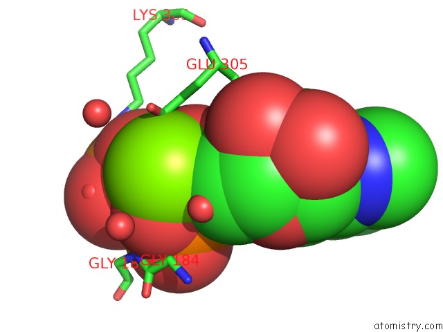
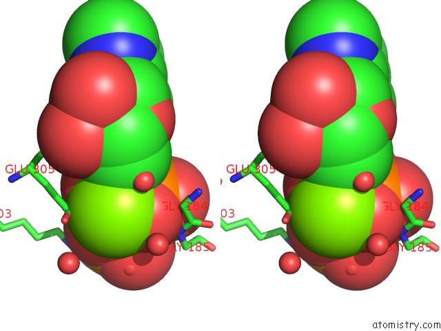
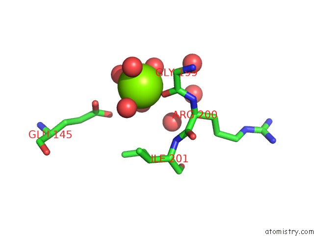
Magnesium binding site 2 out of 6 in 7mxb
Go back to
Magnesium binding site 2 out
of 6 in the Crystal Structure of the S/T Protein Kinase Pkng From Corynebacterium Glutamicum in Complex with Amp-Pnp
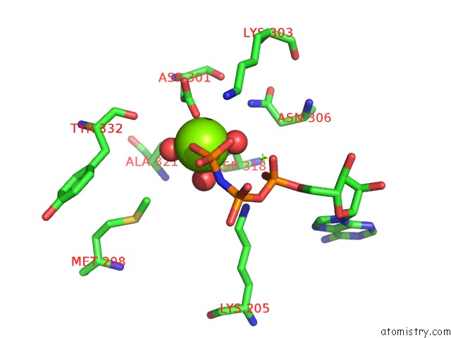
Mono view
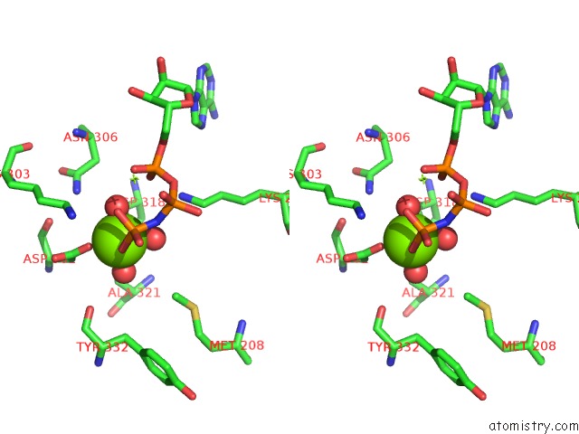
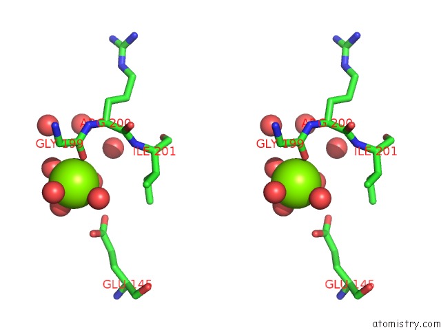
Stereo pair view
Mono view
Stereo pair view
A full contact list of Magnesium with other atoms in the Mg binding
site number 2 of Crystal Structure of the S/T Protein Kinase Pkng From Corynebacterium Glutamicum in Complex with Amp-Pnp within 5.0Å range:
|
Magnesium binding site 3 out of 6 in 7mxb
Go back to
Magnesium binding site 3 out
of 6 in the Crystal Structure of the S/T Protein Kinase Pkng From Corynebacterium Glutamicum in Complex with Amp-Pnp
Mono view
Stereo pair view
Mono view
Stereo pair view
A full contact list of Magnesium with other atoms in the Mg binding
site number 3 of Crystal Structure of the S/T Protein Kinase Pkng From Corynebacterium Glutamicum in Complex with Amp-Pnp within 5.0Å range:
|
Magnesium binding site 4 out of 6 in 7mxb
Go back to
Magnesium binding site 4 out
of 6 in the Crystal Structure of the S/T Protein Kinase Pkng From Corynebacterium Glutamicum in Complex with Amp-Pnp
Mono view
Stereo pair view
Mono view
Stereo pair view
A full contact list of Magnesium with other atoms in the Mg binding
site number 4 of Crystal Structure of the S/T Protein Kinase Pkng From Corynebacterium Glutamicum in Complex with Amp-Pnp within 5.0Å range:
|
Magnesium binding site 5 out of 6 in 7mxb
Go back to
Magnesium binding site 5 out
of 6 in the Crystal Structure of the S/T Protein Kinase Pkng From Corynebacterium Glutamicum in Complex with Amp-Pnp
Mono view
Stereo pair view
Mono view
Stereo pair view
A full contact list of Magnesium with other atoms in the Mg binding
site number 5 of Crystal Structure of the S/T Protein Kinase Pkng From Corynebacterium Glutamicum in Complex with Amp-Pnp within 5.0Å range:
|
Magnesium binding site 6 out of 6 in 7mxb
Go back to
Magnesium binding site 6 out
of 6 in the Crystal Structure of the S/T Protein Kinase Pkng From Corynebacterium Glutamicum in Complex with Amp-Pnp
Mono view
Stereo pair view
Mono view
Stereo pair view
A full contact list of Magnesium with other atoms in the Mg binding
site number 6 of Crystal Structure of the S/T Protein Kinase Pkng From Corynebacterium Glutamicum in Complex with Amp-Pnp within 5.0Å range:
|
Reference:
M.N.Lisa,
A.Sogues,
N.Barilone,
M.Baumgart,
M.Gil,
M.Grana,
R.Duran,
R.M.Biondi,
M.Bellinzoni,
M.Bott,
P.M.Alzari.
A Tetratricopeptide Repeat Scaffold Couples Signal Detection to Odhi Phosphorylation in Metabolic Control By the Protein Kinase Pkng Mbio 2021.
ISSN: ESSN 2150-7511
PubMed: 34607462
DOI: 10.1128/MBIO.01717-21
Page generated: Thu Oct 3 01:09:03 2024
ISSN: ESSN 2150-7511
PubMed: 34607462
DOI: 10.1128/MBIO.01717-21
Last articles
Zn in 9MJ5Zn in 9HNW
Zn in 9G0L
Zn in 9FNE
Zn in 9DZN
Zn in 9E0I
Zn in 9D32
Zn in 9DAK
Zn in 8ZXC
Zn in 8ZUF