Magnesium »
PDB 7nem-7noz »
7nix »
Magnesium in PDB 7nix: 14-3-3 Sigma with AS160 Binding Site PT642
Protein crystallography data
The structure of 14-3-3 Sigma with AS160 Binding Site PT642, PDB code: 7nix
was solved by
M.Wolter,
C.Ottmann,
with X-Ray Crystallography technique. A brief refinement statistics is given in the table below:
| Resolution Low / High (Å) | 60.61 / 1.90 |
| Space group | P 61 2 2 |
| Cell size a, b, c (Å), α, β, γ (°) | 121.219, 121.219, 74.464, 90, 90, 120 |
| R / Rfree (%) | 18.6 / 20.4 |
Magnesium Binding Sites:
The binding sites of Magnesium atom in the 14-3-3 Sigma with AS160 Binding Site PT642
(pdb code 7nix). This binding sites where shown within
5.0 Angstroms radius around Magnesium atom.
In total 2 binding sites of Magnesium where determined in the 14-3-3 Sigma with AS160 Binding Site PT642, PDB code: 7nix:
Jump to Magnesium binding site number: 1; 2;
In total 2 binding sites of Magnesium where determined in the 14-3-3 Sigma with AS160 Binding Site PT642, PDB code: 7nix:
Jump to Magnesium binding site number: 1; 2;
Magnesium binding site 1 out of 2 in 7nix
Go back to
Magnesium binding site 1 out
of 2 in the 14-3-3 Sigma with AS160 Binding Site PT642
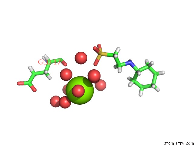
Mono view
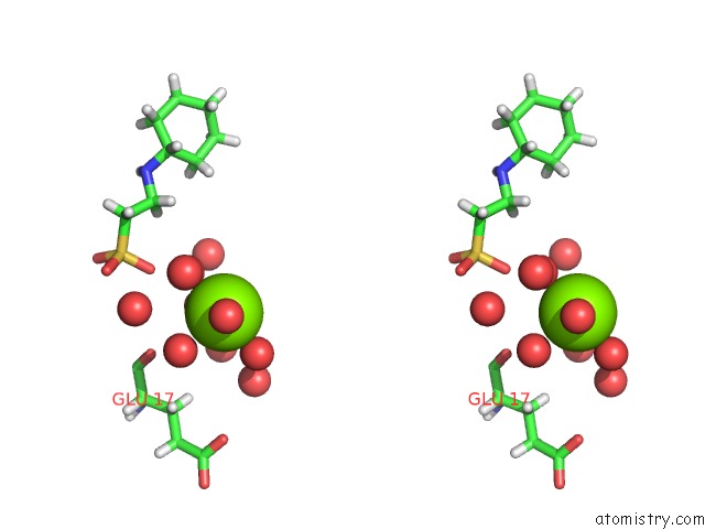
Stereo pair view
Mono view
Stereo pair view
A full contact list of Magnesium with other atoms in the Mg binding
site number 1 of 14-3-3 Sigma with AS160 Binding Site PT642 within 5.0Å range:
|
Magnesium binding site 2 out of 2 in 7nix
Go back to
Magnesium binding site 2 out
of 2 in the 14-3-3 Sigma with AS160 Binding Site PT642
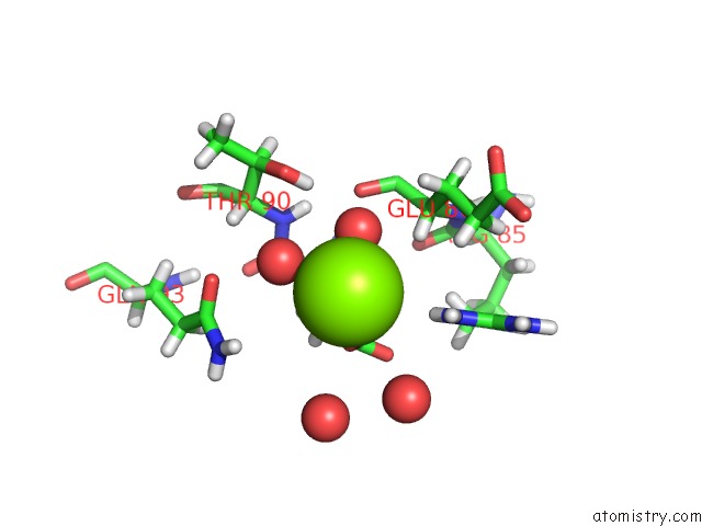
Mono view
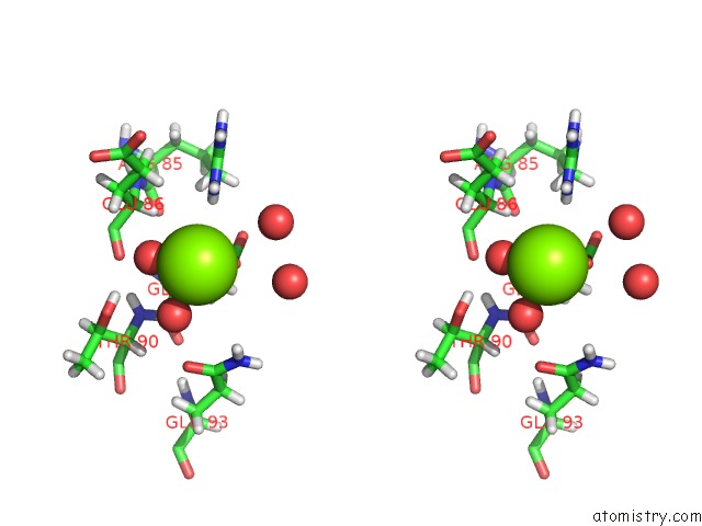
Stereo pair view
Mono view
Stereo pair view
A full contact list of Magnesium with other atoms in the Mg binding
site number 2 of 14-3-3 Sigma with AS160 Binding Site PT642 within 5.0Å range:
|
Reference:
P.J.Cossar,
M.Wolter,
L.Van Dijck,
D.Valenti,
L.M.Levy,
C.Ottmann,
L.Brunsveld.
Reversible Covalent Imine-Tethering For Selective Stabilization of 14-3-3 Hub Protein Interactions. J.Am.Chem.Soc. 2021.
ISSN: ESSN 1520-5126
PubMed: 34047554
DOI: 10.1021/JACS.1C03035
Page generated: Thu Oct 3 02:05:03 2024
ISSN: ESSN 1520-5126
PubMed: 34047554
DOI: 10.1021/JACS.1C03035
Last articles
Zn in 9MJ5Zn in 9HNW
Zn in 9G0L
Zn in 9FNE
Zn in 9DZN
Zn in 9E0I
Zn in 9D32
Zn in 9DAK
Zn in 8ZXC
Zn in 8ZUF