Magnesium »
PDB 7np2-7nvm »
7nsr »
Magnesium in PDB 7nsr: Myelin Protein P2 I50DEL
Protein crystallography data
The structure of Myelin Protein P2 I50DEL, PDB code: 7nsr
was solved by
M.Uusitalo,
S.Ruskamo,
P.Kursula,
with X-Ray Crystallography technique. A brief refinement statistics is given in the table below:
| Resolution Low / High (Å) | 46.77 / 1.50 |
| Space group | P 41 21 2 |
| Cell size a, b, c (Å), α, β, γ (°) | 66.14, 66.14, 101.19, 90, 90, 90 |
| R / Rfree (%) | 18.2 / 19.9 |
Other elements in 7nsr:
The structure of Myelin Protein P2 I50DEL also contains other interesting chemical elements:
| Chlorine | (Cl) | 1 atom |
Magnesium Binding Sites:
The binding sites of Magnesium atom in the Myelin Protein P2 I50DEL
(pdb code 7nsr). This binding sites where shown within
5.0 Angstroms radius around Magnesium atom.
In total 2 binding sites of Magnesium where determined in the Myelin Protein P2 I50DEL, PDB code: 7nsr:
Jump to Magnesium binding site number: 1; 2;
In total 2 binding sites of Magnesium where determined in the Myelin Protein P2 I50DEL, PDB code: 7nsr:
Jump to Magnesium binding site number: 1; 2;
Magnesium binding site 1 out of 2 in 7nsr
Go back to
Magnesium binding site 1 out
of 2 in the Myelin Protein P2 I50DEL
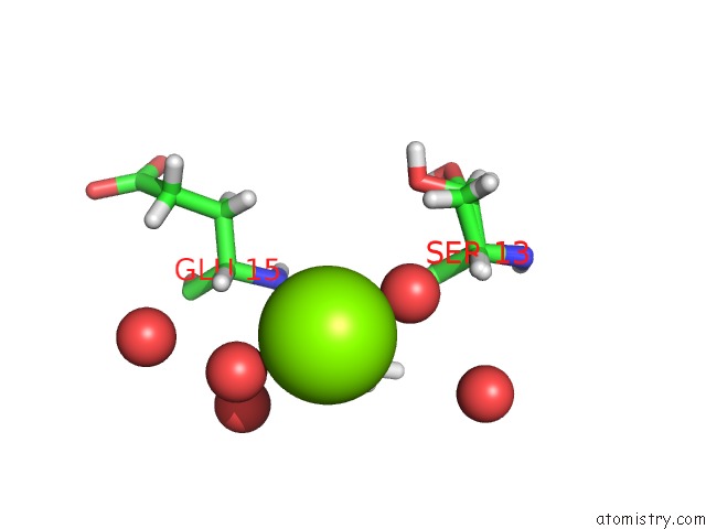
Mono view
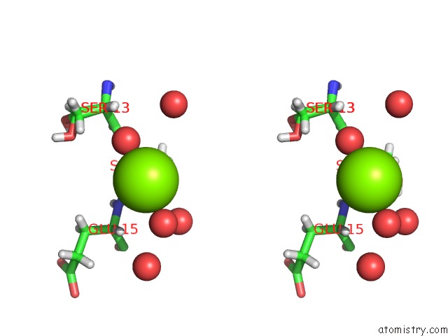
Stereo pair view
Mono view
Stereo pair view
A full contact list of Magnesium with other atoms in the Mg binding
site number 1 of Myelin Protein P2 I50DEL within 5.0Å range:
|
Magnesium binding site 2 out of 2 in 7nsr
Go back to
Magnesium binding site 2 out
of 2 in the Myelin Protein P2 I50DEL
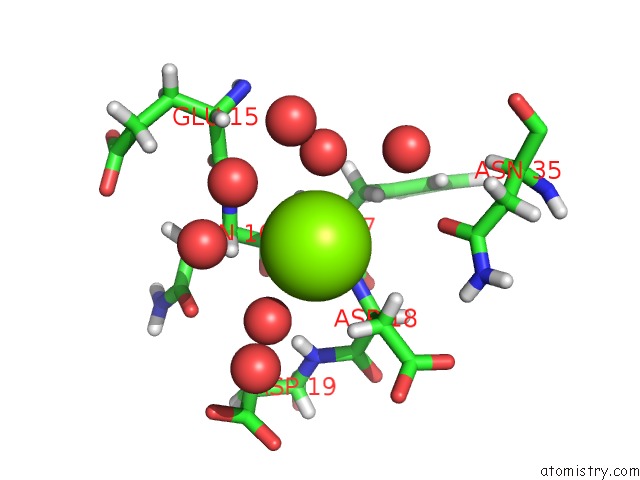
Mono view
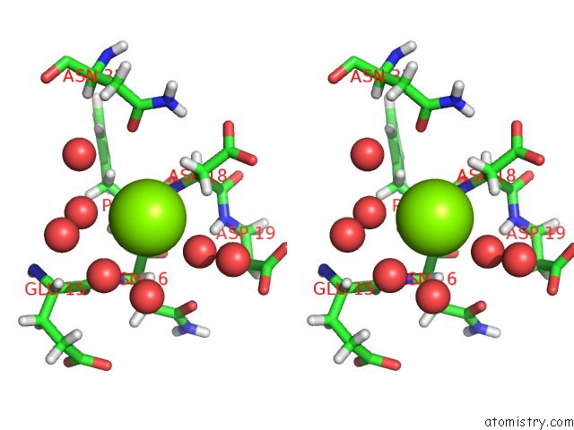
Stereo pair view
Mono view
Stereo pair view
A full contact list of Magnesium with other atoms in the Mg binding
site number 2 of Myelin Protein P2 I50DEL within 5.0Å range:
|
Reference:
M.Uusitalo,
M.B.Klenow,
S.Laulumaa,
M.P.Blakeley,
A.C.Simonsen,
S.Ruskamo,
P.Kursula.
Human Myelin Protein P2: From Crystallography to Time-Lapse Membrane Imaging and Neuropathy-Associated Variants. Febs J. 2021.
ISSN: ISSN 1742-464X
PubMed: 34138518
DOI: 10.1111/FEBS.16079
Page generated: Thu Oct 3 02:13:11 2024
ISSN: ISSN 1742-464X
PubMed: 34138518
DOI: 10.1111/FEBS.16079
Last articles
Fe in 2YXOFe in 2YRS
Fe in 2YXC
Fe in 2YNM
Fe in 2YVJ
Fe in 2YP1
Fe in 2YU2
Fe in 2YU1
Fe in 2YQB
Fe in 2YOO