Magnesium »
PDB 7o6c-7ocq »
7obc »
Magnesium in PDB 7obc: Crystal Structure of 14-3-3 Sigma in Complex with Phosphorylated and Farnesylated RND3 Peptide
Protein crystallography data
The structure of Crystal Structure of 14-3-3 Sigma in Complex with Phosphorylated and Farnesylated RND3 Peptide, PDB code: 7obc
was solved by
F.Centorrino,
B.Andlovic,
C.Ottmann,
with X-Ray Crystallography technique. A brief refinement statistics is given in the table below:
| Resolution Low / High (Å) | 29.90 / 1.90 |
| Space group | C 2 2 21 |
| Cell size a, b, c (Å), α, β, γ (°) | 82.4, 111.95, 62.886, 90, 90, 90 |
| R / Rfree (%) | 15.1 / 20 |
Other elements in 7obc:
The structure of Crystal Structure of 14-3-3 Sigma in Complex with Phosphorylated and Farnesylated RND3 Peptide also contains other interesting chemical elements:
| Chlorine | (Cl) | 1 atom |
Magnesium Binding Sites:
The binding sites of Magnesium atom in the Crystal Structure of 14-3-3 Sigma in Complex with Phosphorylated and Farnesylated RND3 Peptide
(pdb code 7obc). This binding sites where shown within
5.0 Angstroms radius around Magnesium atom.
In total only one binding site of Magnesium was determined in the Crystal Structure of 14-3-3 Sigma in Complex with Phosphorylated and Farnesylated RND3 Peptide, PDB code: 7obc:
In total only one binding site of Magnesium was determined in the Crystal Structure of 14-3-3 Sigma in Complex with Phosphorylated and Farnesylated RND3 Peptide, PDB code: 7obc:
Magnesium binding site 1 out of 1 in 7obc
Go back to
Magnesium binding site 1 out
of 1 in the Crystal Structure of 14-3-3 Sigma in Complex with Phosphorylated and Farnesylated RND3 Peptide

Mono view
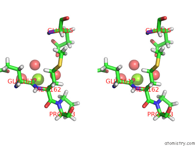
Stereo pair view
Mono view
Stereo pair view
A full contact list of Magnesium with other atoms in the Mg binding
site number 1 of Crystal Structure of 14-3-3 Sigma in Complex with Phosphorylated and Farnesylated RND3 Peptide within 5.0Å range:
|
Reference:
B.Andlovic,
G.Heilmann,
S.Ninck,
S.Andrei,
F.Centorrino,
Y.Higuchi,
N.Kato,
L.Brunsveld,
S.Menninger,
A.Choidas,
A.Wolf,
M.Kaiser,
J.Eickhoff,
C.Ottmann.
Inf Alpha Primes Ovarian Cancer Cells For Fusicoccin-Induced Cell Death Via Stabilization of 14-3-3 Protein-Protein Interactions To Be Published.
Page generated: Thu Oct 3 02:52:21 2024
Last articles
Zn in 9MJ5Zn in 9HNW
Zn in 9G0L
Zn in 9FNE
Zn in 9DZN
Zn in 9E0I
Zn in 9D32
Zn in 9DAK
Zn in 8ZXC
Zn in 8ZUF