Magnesium »
PDB 7oup-7p8o »
7oyf »
Magnesium in PDB 7oyf: Crystal Structure of Depupylase Dop in Complex with Pup and Adp/Trifluoromagnesate
Protein crystallography data
The structure of Crystal Structure of Depupylase Dop in Complex with Pup and Adp/Trifluoromagnesate, PDB code: 7oyf
was solved by
H.Cui,
with X-Ray Crystallography technique. A brief refinement statistics is given in the table below:
| Resolution Low / High (Å) | 47.65 / 1.88 |
| Space group | P 31 2 1 |
| Cell size a, b, c (Å), α, β, γ (°) | 106.334, 106.334, 107.384, 90, 90, 120 |
| R / Rfree (%) | 15.9 / 18.8 |
Other elements in 7oyf:
The structure of Crystal Structure of Depupylase Dop in Complex with Pup and Adp/Trifluoromagnesate also contains other interesting chemical elements:
| Fluorine | (F) | 3 atoms |
Magnesium Binding Sites:
The binding sites of Magnesium atom in the Crystal Structure of Depupylase Dop in Complex with Pup and Adp/Trifluoromagnesate
(pdb code 7oyf). This binding sites where shown within
5.0 Angstroms radius around Magnesium atom.
In total 4 binding sites of Magnesium where determined in the Crystal Structure of Depupylase Dop in Complex with Pup and Adp/Trifluoromagnesate, PDB code: 7oyf:
Jump to Magnesium binding site number: 1; 2; 3; 4;
In total 4 binding sites of Magnesium where determined in the Crystal Structure of Depupylase Dop in Complex with Pup and Adp/Trifluoromagnesate, PDB code: 7oyf:
Jump to Magnesium binding site number: 1; 2; 3; 4;
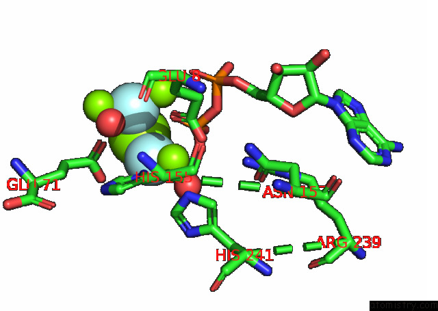
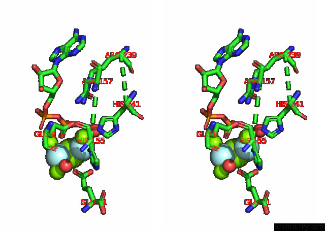
Magnesium binding site 1 out of 4 in 7oyf
Go back to
Magnesium binding site 1 out
of 4 in the Crystal Structure of Depupylase Dop in Complex with Pup and Adp/Trifluoromagnesate
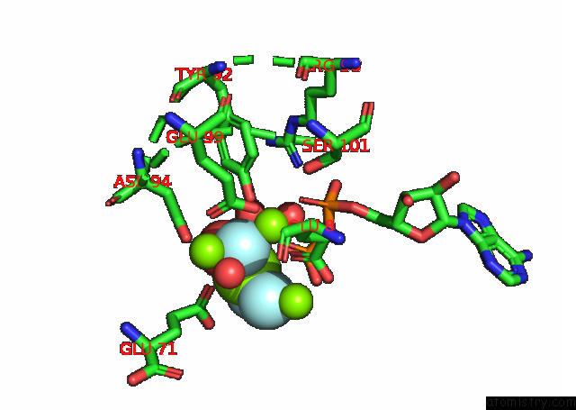
Mono view
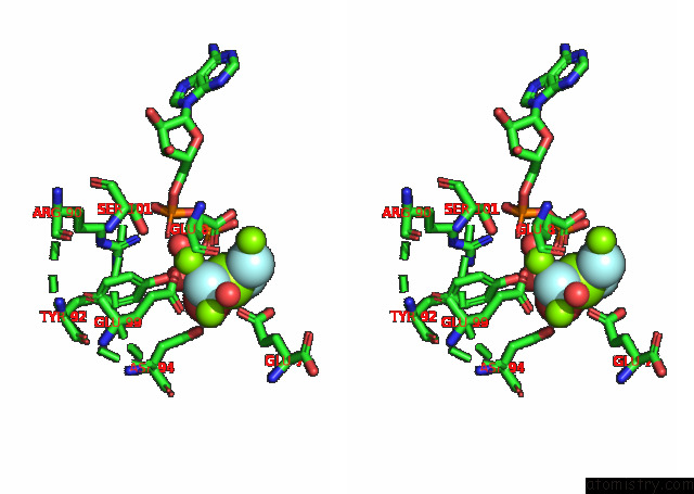
Stereo pair view
Mono view
Stereo pair view
A full contact list of Magnesium with other atoms in the Mg binding
site number 1 of Crystal Structure of Depupylase Dop in Complex with Pup and Adp/Trifluoromagnesate within 5.0Å range:
|
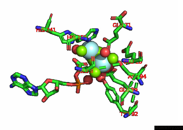
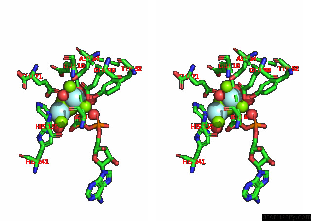
Magnesium binding site 2 out of 4 in 7oyf
Go back to
Magnesium binding site 2 out
of 4 in the Crystal Structure of Depupylase Dop in Complex with Pup and Adp/Trifluoromagnesate
Mono view
Stereo pair view
Mono view
Stereo pair view
A full contact list of Magnesium with other atoms in the Mg binding
site number 2 of Crystal Structure of Depupylase Dop in Complex with Pup and Adp/Trifluoromagnesate within 5.0Å range:
|
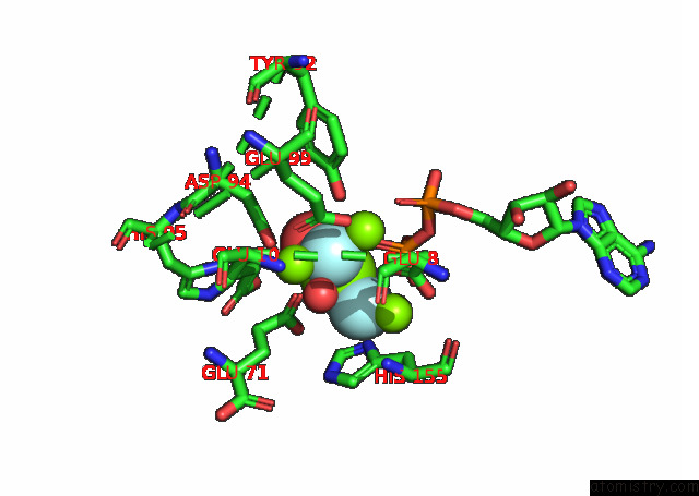
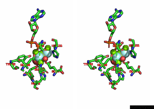
Magnesium binding site 3 out of 4 in 7oyf
Go back to
Magnesium binding site 3 out
of 4 in the Crystal Structure of Depupylase Dop in Complex with Pup and Adp/Trifluoromagnesate
Mono view
Stereo pair view
Mono view
Stereo pair view
A full contact list of Magnesium with other atoms in the Mg binding
site number 3 of Crystal Structure of Depupylase Dop in Complex with Pup and Adp/Trifluoromagnesate within 5.0Å range:
|
Magnesium binding site 4 out of 4 in 7oyf
Go back to
Magnesium binding site 4 out
of 4 in the Crystal Structure of Depupylase Dop in Complex with Pup and Adp/Trifluoromagnesate
Mono view
Stereo pair view
Mono view
Stereo pair view
A full contact list of Magnesium with other atoms in the Mg binding
site number 4 of Crystal Structure of Depupylase Dop in Complex with Pup and Adp/Trifluoromagnesate within 5.0Å range:
|
Reference:
H.Cui,
A.U.Muller,
M.Leibundgut,
J.Tian,
N.Ban,
E.Weber-Ban.
Structures of Prokaryotic Ubiquitin-Like Protein Pup in Complex with Depupylase Dop Reveal the Mechanism of Catalytic Phosphate Formation. Nat Commun V. 12 6635 2021.
ISSN: ESSN 2041-1723
PubMed: 34789727
DOI: 10.1038/S41467-021-26848-X
Page generated: Thu Aug 14 12:39:48 2025
ISSN: ESSN 2041-1723
PubMed: 34789727
DOI: 10.1038/S41467-021-26848-X
Last articles
Mg in 7R8IMg in 7R8E
Mg in 7R7J
Mg in 7R7G
Mg in 7R7F
Mg in 7R6Y
Mg in 7R7E
Mg in 7R78
Mg in 7R6V
Mg in 7R6N