Magnesium »
PDB 7oug-7p84 »
7p0q »
Magnesium in PDB 7p0q: F(M197)H Mutant Structure of Photosynthetic Reaction Center From Rhodobacter Sphaeroides Strain Rv By Fixed-Target Serial Synchrotron Crystallography (100K, 26KEV)
Protein crystallography data
The structure of F(M197)H Mutant Structure of Photosynthetic Reaction Center From Rhodobacter Sphaeroides Strain Rv By Fixed-Target Serial Synchrotron Crystallography (100K, 26KEV), PDB code: 7p0q
was solved by
A.G.Gabdulkhakov,
G.K.Selikhanov,
S.Guenther,
A.Meents,
T.Y.Fufina,
L.G.Vasilieva,
with X-Ray Crystallography technique. A brief refinement statistics is given in the table below:
| Resolution Low / High (Å) | 48.93 / 1.73 |
| Space group | P 42 21 2 |
| Cell size a, b, c (Å), α, β, γ (°) | 99.97, 99.97, 239.22, 90, 90, 90 |
| R / Rfree (%) | 18.2 / 20.3 |
Other elements in 7p0q:
The structure of F(M197)H Mutant Structure of Photosynthetic Reaction Center From Rhodobacter Sphaeroides Strain Rv By Fixed-Target Serial Synchrotron Crystallography (100K, 26KEV) also contains other interesting chemical elements:
| Iron | (Fe) | 1 atom |
Magnesium Binding Sites:
The binding sites of Magnesium atom in the F(M197)H Mutant Structure of Photosynthetic Reaction Center From Rhodobacter Sphaeroides Strain Rv By Fixed-Target Serial Synchrotron Crystallography (100K, 26KEV)
(pdb code 7p0q). This binding sites where shown within
5.0 Angstroms radius around Magnesium atom.
In total 4 binding sites of Magnesium where determined in the F(M197)H Mutant Structure of Photosynthetic Reaction Center From Rhodobacter Sphaeroides Strain Rv By Fixed-Target Serial Synchrotron Crystallography (100K, 26KEV), PDB code: 7p0q:
Jump to Magnesium binding site number: 1; 2; 3; 4;
In total 4 binding sites of Magnesium where determined in the F(M197)H Mutant Structure of Photosynthetic Reaction Center From Rhodobacter Sphaeroides Strain Rv By Fixed-Target Serial Synchrotron Crystallography (100K, 26KEV), PDB code: 7p0q:
Jump to Magnesium binding site number: 1; 2; 3; 4;
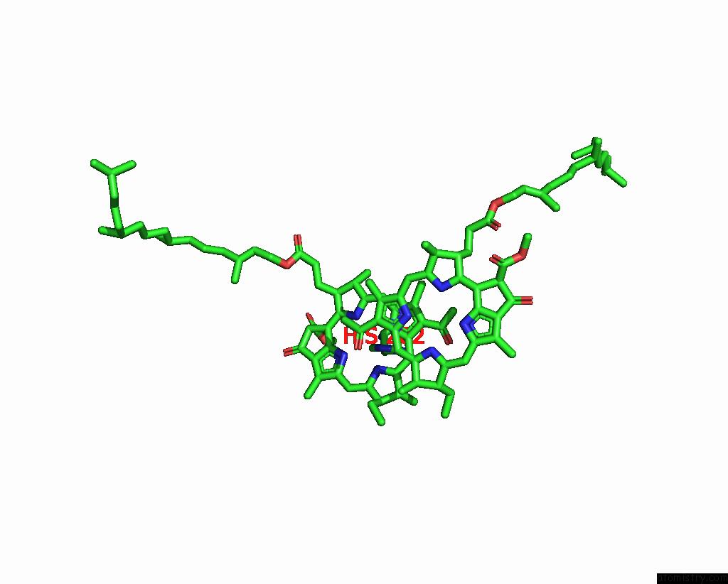
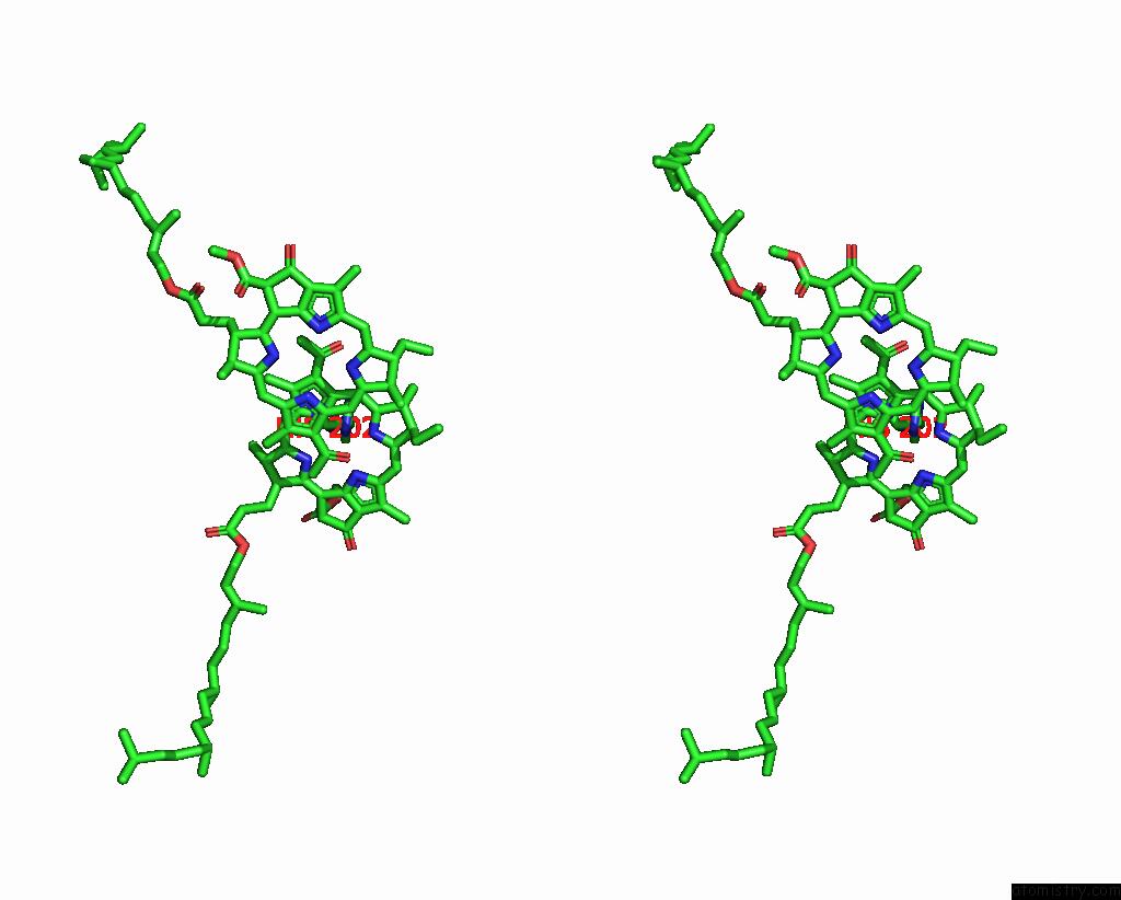
Magnesium binding site 1 out of 4 in 7p0q
Go back to
Magnesium binding site 1 out
of 4 in the F(M197)H Mutant Structure of Photosynthetic Reaction Center From Rhodobacter Sphaeroides Strain Rv By Fixed-Target Serial Synchrotron Crystallography (100K, 26KEV)
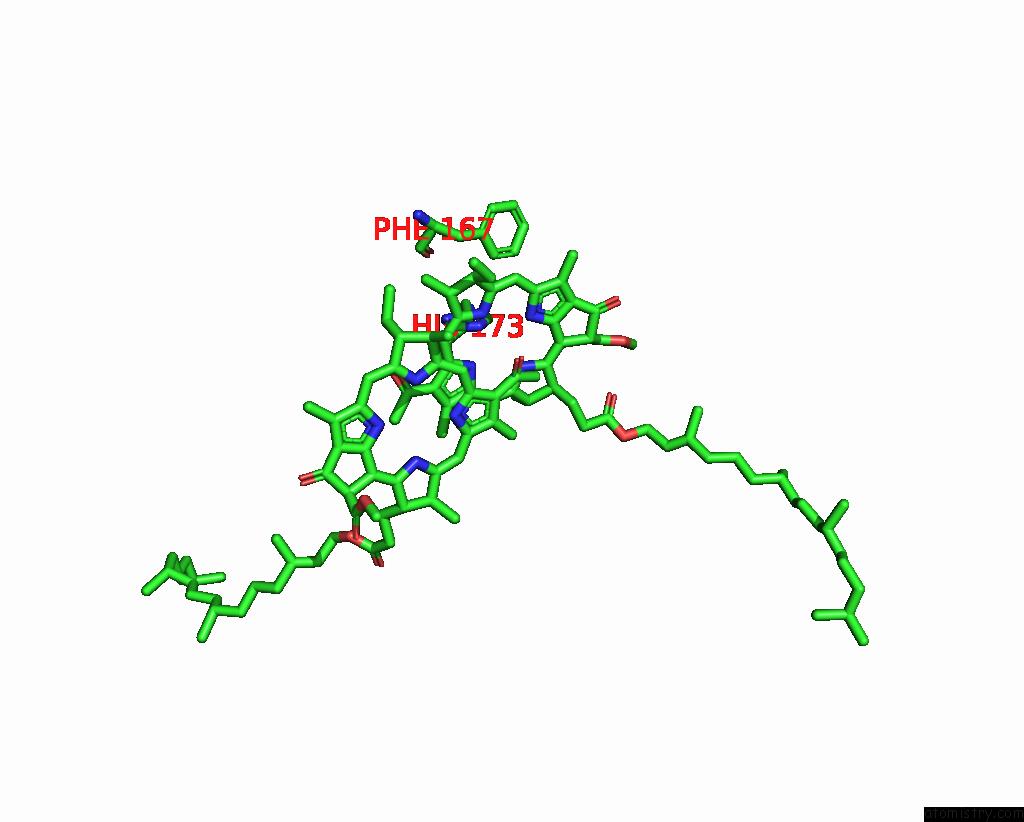
Mono view
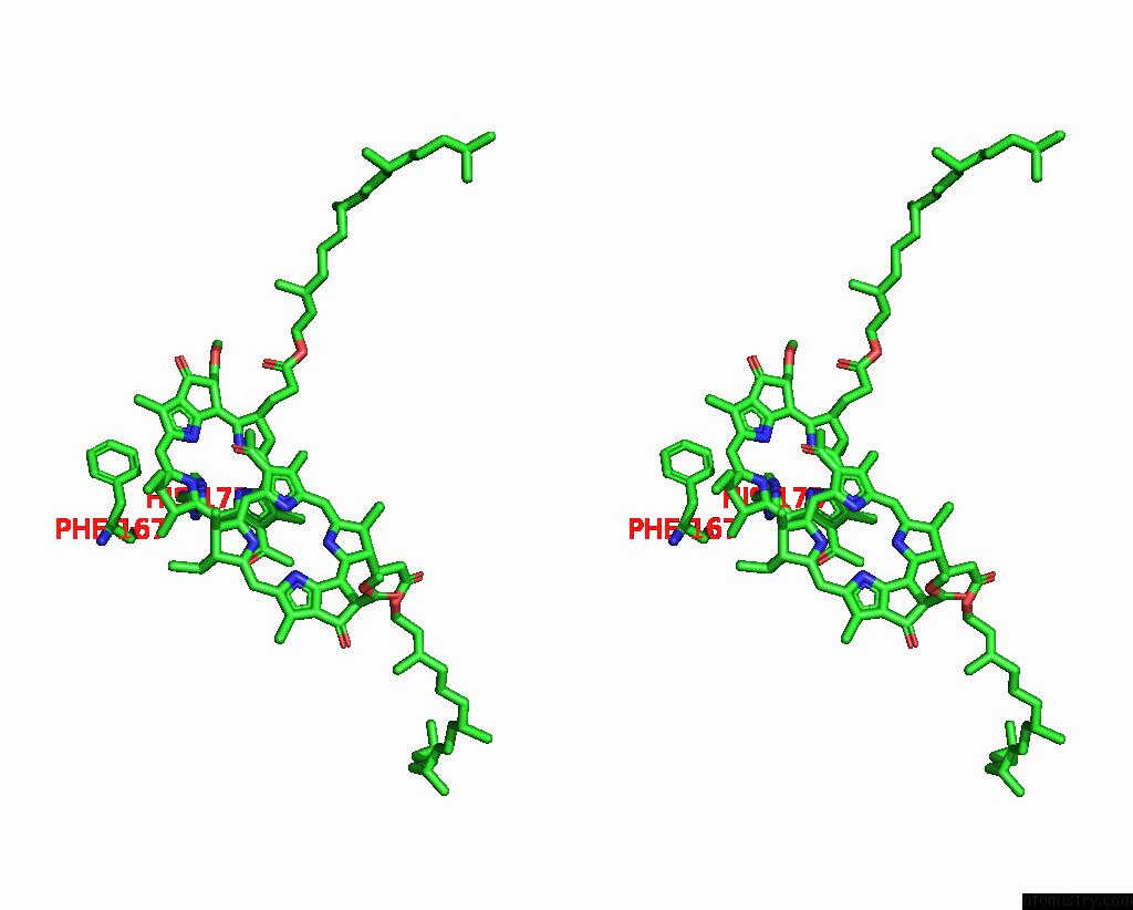
Stereo pair view
Mono view
Stereo pair view
A full contact list of Magnesium with other atoms in the Mg binding
site number 1 of F(M197)H Mutant Structure of Photosynthetic Reaction Center From Rhodobacter Sphaeroides Strain Rv By Fixed-Target Serial Synchrotron Crystallography (100K, 26KEV) within 5.0Å range:
|
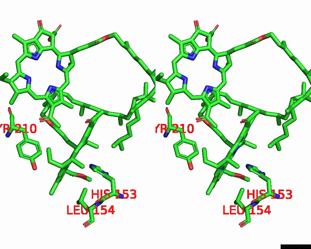
Magnesium binding site 2 out of 4 in 7p0q
Go back to
Magnesium binding site 2 out
of 4 in the F(M197)H Mutant Structure of Photosynthetic Reaction Center From Rhodobacter Sphaeroides Strain Rv By Fixed-Target Serial Synchrotron Crystallography (100K, 26KEV)

Mono view
Stereo pair view
Mono view
Stereo pair view
A full contact list of Magnesium with other atoms in the Mg binding
site number 2 of F(M197)H Mutant Structure of Photosynthetic Reaction Center From Rhodobacter Sphaeroides Strain Rv By Fixed-Target Serial Synchrotron Crystallography (100K, 26KEV) within 5.0Å range:
|
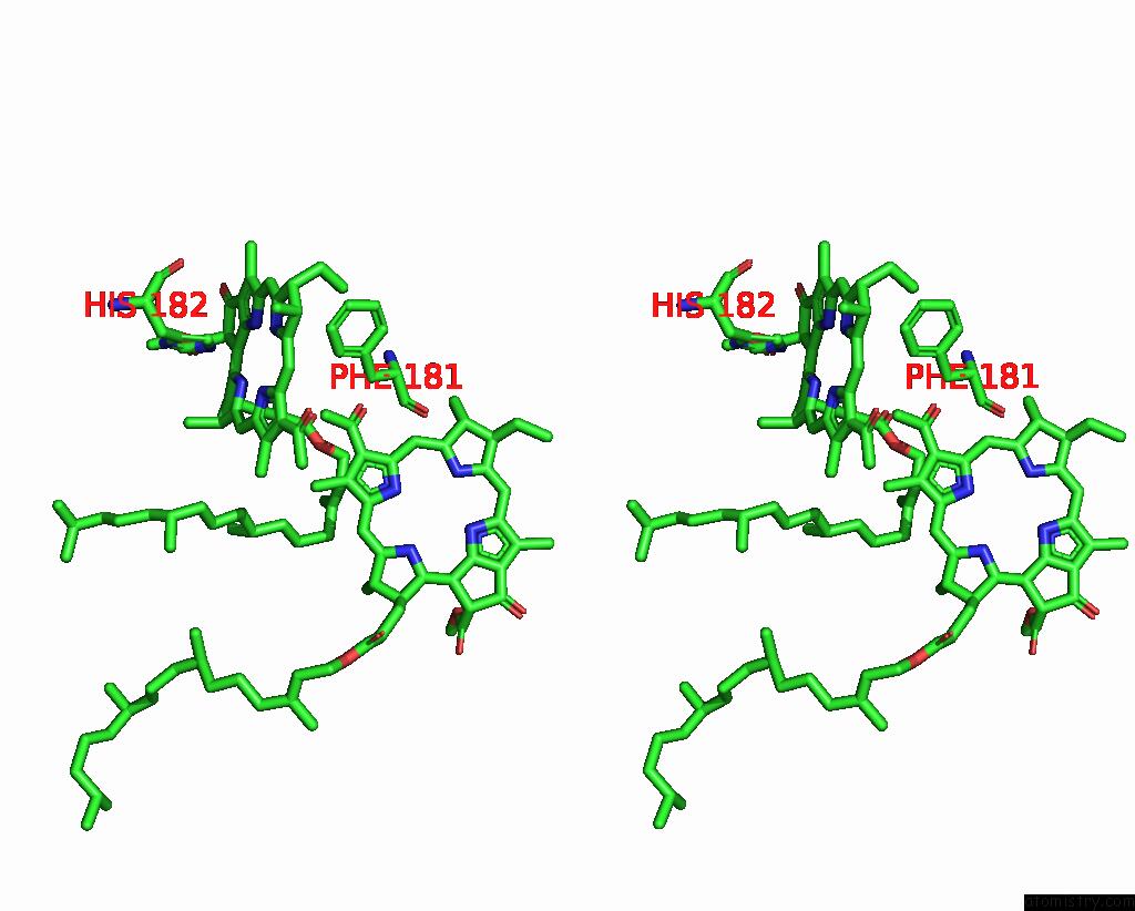
Magnesium binding site 3 out of 4 in 7p0q
Go back to
Magnesium binding site 3 out
of 4 in the F(M197)H Mutant Structure of Photosynthetic Reaction Center From Rhodobacter Sphaeroides Strain Rv By Fixed-Target Serial Synchrotron Crystallography (100K, 26KEV)

Mono view
Stereo pair view
Mono view
Stereo pair view
A full contact list of Magnesium with other atoms in the Mg binding
site number 3 of F(M197)H Mutant Structure of Photosynthetic Reaction Center From Rhodobacter Sphaeroides Strain Rv By Fixed-Target Serial Synchrotron Crystallography (100K, 26KEV) within 5.0Å range:
|
Magnesium binding site 4 out of 4 in 7p0q
Go back to
Magnesium binding site 4 out
of 4 in the F(M197)H Mutant Structure of Photosynthetic Reaction Center From Rhodobacter Sphaeroides Strain Rv By Fixed-Target Serial Synchrotron Crystallography (100K, 26KEV)
Mono view
Stereo pair view
Mono view
Stereo pair view
A full contact list of Magnesium with other atoms in the Mg binding
site number 4 of F(M197)H Mutant Structure of Photosynthetic Reaction Center From Rhodobacter Sphaeroides Strain Rv By Fixed-Target Serial Synchrotron Crystallography (100K, 26KEV) within 5.0Å range:
|
Reference:
A.G.Gabdulkhakov,
G.K.Selikhanov,
S.Guenther,
A.Meents,
T.Y.Fufina,
L.G.Vasilieva.
X-Ray Structure of Rhodobacter Sphaeroides Reaction Center with M197 Phe-His Substitution Clarifies Properties of the Mutant Complex To Be Published.
Page generated: Thu Oct 3 03:58:05 2024
Last articles
Zn in 9MJ5Zn in 9HNW
Zn in 9G0L
Zn in 9FNE
Zn in 9DZN
Zn in 9E0I
Zn in 9D32
Zn in 9DAK
Zn in 8ZXC
Zn in 8ZUF