Magnesium »
PDB 7p8d-7pje »
7pe4 »
Magnesium in PDB 7pe4: Crystal Structure of Mycobacterium Hassiacum Glucosyl-3- Phosphoglycerate Synthase at pH 5.5 in Complex with Udp-Glucose
Enzymatic activity of Crystal Structure of Mycobacterium Hassiacum Glucosyl-3- Phosphoglycerate Synthase at pH 5.5 in Complex with Udp-Glucose
All present enzymatic activity of Crystal Structure of Mycobacterium Hassiacum Glucosyl-3- Phosphoglycerate Synthase at pH 5.5 in Complex with Udp-Glucose:
2.4.1.266;
2.4.1.266;
Protein crystallography data
The structure of Crystal Structure of Mycobacterium Hassiacum Glucosyl-3- Phosphoglycerate Synthase at pH 5.5 in Complex with Udp-Glucose, PDB code: 7pe4
was solved by
A.Silva,
P.J.Babosa Pereira,
S.Macedo-Ribeiro,
with X-Ray Crystallography technique. A brief refinement statistics is given in the table below:
| Resolution Low / High (Å) | 46.58 / 2.05 |
| Space group | I 41 |
| Cell size a, b, c (Å), α, β, γ (°) | 101.887, 101.887, 122.098, 90, 90, 90 |
| R / Rfree (%) | 16 / 19.2 |
Other elements in 7pe4:
The structure of Crystal Structure of Mycobacterium Hassiacum Glucosyl-3- Phosphoglycerate Synthase at pH 5.5 in Complex with Udp-Glucose also contains other interesting chemical elements:
| Chlorine | (Cl) | 3 atoms |
Magnesium Binding Sites:
The binding sites of Magnesium atom in the Crystal Structure of Mycobacterium Hassiacum Glucosyl-3- Phosphoglycerate Synthase at pH 5.5 in Complex with Udp-Glucose
(pdb code 7pe4). This binding sites where shown within
5.0 Angstroms radius around Magnesium atom.
In total only one binding site of Magnesium was determined in the Crystal Structure of Mycobacterium Hassiacum Glucosyl-3- Phosphoglycerate Synthase at pH 5.5 in Complex with Udp-Glucose, PDB code: 7pe4:
In total only one binding site of Magnesium was determined in the Crystal Structure of Mycobacterium Hassiacum Glucosyl-3- Phosphoglycerate Synthase at pH 5.5 in Complex with Udp-Glucose, PDB code: 7pe4:
Magnesium binding site 1 out of 1 in 7pe4
Go back to
Magnesium binding site 1 out
of 1 in the Crystal Structure of Mycobacterium Hassiacum Glucosyl-3- Phosphoglycerate Synthase at pH 5.5 in Complex with Udp-Glucose
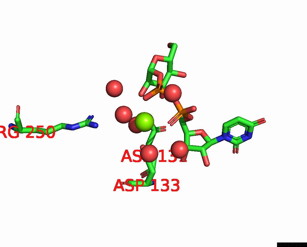
Mono view
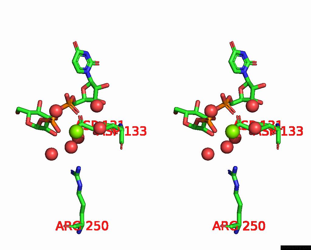
Stereo pair view
Mono view
Stereo pair view
A full contact list of Magnesium with other atoms in the Mg binding
site number 1 of Crystal Structure of Mycobacterium Hassiacum Glucosyl-3- Phosphoglycerate Synthase at pH 5.5 in Complex with Udp-Glucose within 5.0Å range:
|
Reference:
A.Silva,
D.Nunes-Costa,
N.Empadinhas,
P.J.Babosa Pereira,
S.Macedo-Ribeiro.
Crystal Structure of Mycobacterium Hassiacum Glucosyl-3-Phosphoglycerate Synthase at pH 5.5 in Complex with Udp-Glucose To Be Published.
Page generated: Thu Oct 3 04:09:26 2024
Last articles
Fe in 2YXOFe in 2YRS
Fe in 2YXC
Fe in 2YNM
Fe in 2YVJ
Fe in 2YP1
Fe in 2YU2
Fe in 2YU1
Fe in 2YQB
Fe in 2YOO