Magnesium »
PDB 7pn9-7py0 »
7pvk »
Magnesium in PDB 7pvk: X-Ray Structure of Dimeric Porx (T272A Mutant), in Complex with Pgpg.
Protein crystallography data
The structure of X-Ray Structure of Dimeric Porx (T272A Mutant), in Complex with Pgpg., PDB code: 7pvk
was solved by
C.A.Schmitz,
M.Madej,
J.Potempa,
M.Sola,
with X-Ray Crystallography technique. A brief refinement statistics is given in the table below:
| Resolution Low / High (Å) | 51.90 / 2.40 |
| Space group | P 1 21 1 |
| Cell size a, b, c (Å), α, β, γ (°) | 94.63, 103.77, 132.21, 90, 98.5, 90 |
| R / Rfree (%) | 19.4 / 22.3 |
Other elements in 7pvk:
The structure of X-Ray Structure of Dimeric Porx (T272A Mutant), in Complex with Pgpg. also contains other interesting chemical elements:
| Fluorine | (F) | 12 atoms |
| Zinc | (Zn) | 10 atoms |
Magnesium Binding Sites:
The binding sites of Magnesium atom in the X-Ray Structure of Dimeric Porx (T272A Mutant), in Complex with Pgpg.
(pdb code 7pvk). This binding sites where shown within
5.0 Angstroms radius around Magnesium atom.
In total 4 binding sites of Magnesium where determined in the X-Ray Structure of Dimeric Porx (T272A Mutant), in Complex with Pgpg., PDB code: 7pvk:
Jump to Magnesium binding site number: 1; 2; 3; 4;
In total 4 binding sites of Magnesium where determined in the X-Ray Structure of Dimeric Porx (T272A Mutant), in Complex with Pgpg., PDB code: 7pvk:
Jump to Magnesium binding site number: 1; 2; 3; 4;
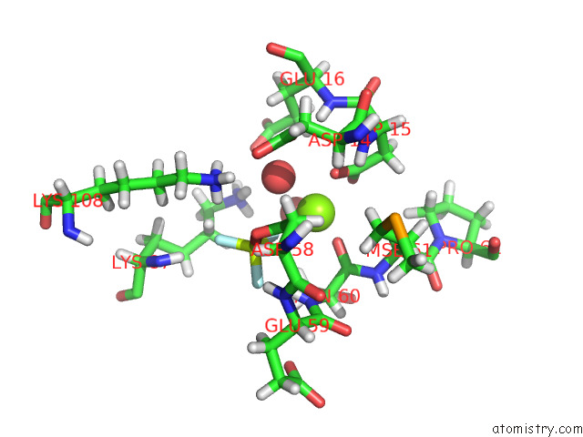
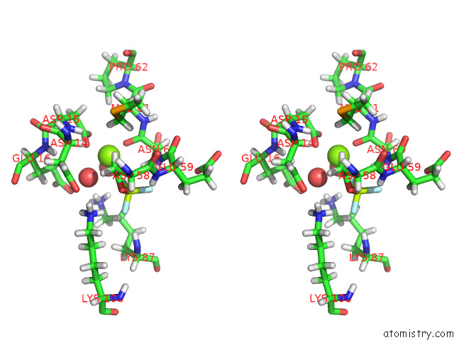
Magnesium binding site 1 out of 4 in 7pvk
Go back to
Magnesium binding site 1 out
of 4 in the X-Ray Structure of Dimeric Porx (T272A Mutant), in Complex with Pgpg.
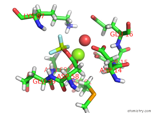
Mono view
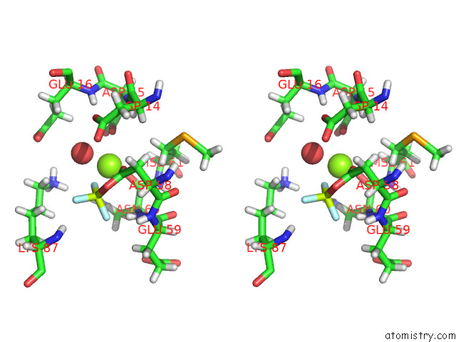
Stereo pair view
Mono view
Stereo pair view
A full contact list of Magnesium with other atoms in the Mg binding
site number 1 of X-Ray Structure of Dimeric Porx (T272A Mutant), in Complex with Pgpg. within 5.0Å range:
|
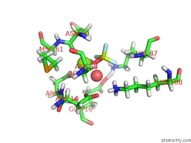
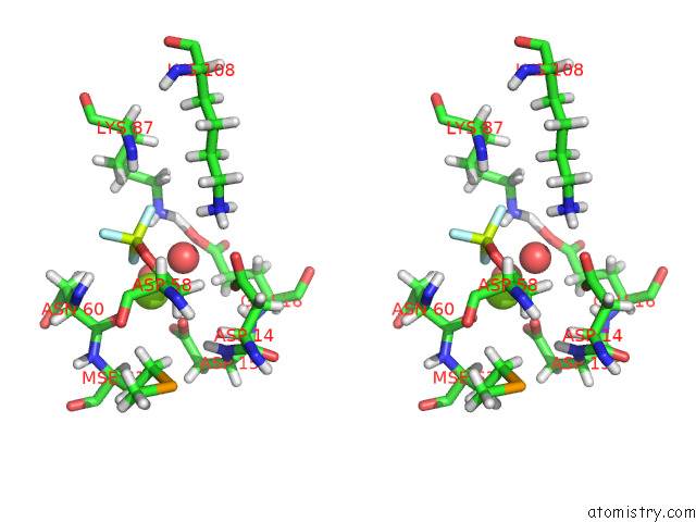
Magnesium binding site 2 out of 4 in 7pvk
Go back to
Magnesium binding site 2 out
of 4 in the X-Ray Structure of Dimeric Porx (T272A Mutant), in Complex with Pgpg.
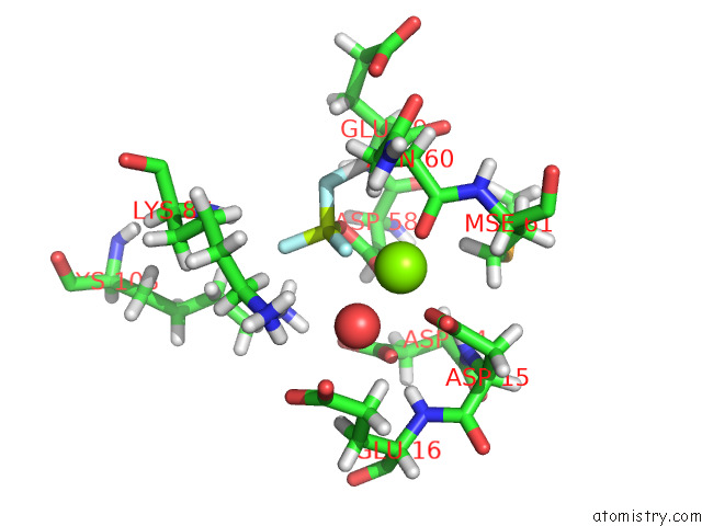
Mono view
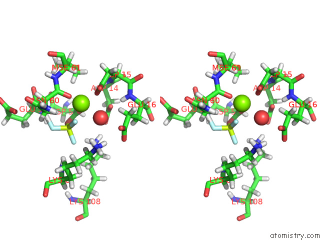
Stereo pair view
Mono view
Stereo pair view
A full contact list of Magnesium with other atoms in the Mg binding
site number 2 of X-Ray Structure of Dimeric Porx (T272A Mutant), in Complex with Pgpg. within 5.0Å range:
|
Magnesium binding site 3 out of 4 in 7pvk
Go back to
Magnesium binding site 3 out
of 4 in the X-Ray Structure of Dimeric Porx (T272A Mutant), in Complex with Pgpg.
Mono view
Stereo pair view
Mono view
Stereo pair view
A full contact list of Magnesium with other atoms in the Mg binding
site number 3 of X-Ray Structure of Dimeric Porx (T272A Mutant), in Complex with Pgpg. within 5.0Å range:
|
Magnesium binding site 4 out of 4 in 7pvk
Go back to
Magnesium binding site 4 out
of 4 in the X-Ray Structure of Dimeric Porx (T272A Mutant), in Complex with Pgpg.
Mono view
Stereo pair view
Mono view
Stereo pair view
A full contact list of Magnesium with other atoms in the Mg binding
site number 4 of X-Ray Structure of Dimeric Porx (T272A Mutant), in Complex with Pgpg. within 5.0Å range:
|
Reference:
C.Schmitz,
M.Madej,
Z.Nowakowska,
A.Cuppari,
A.Jacula,
M.Ksiazek,
K.Mikruta,
J.Wisniewski,
N.Pudelko-Malik,
A.Saran,
N.Zeytuni,
P.Mlynarz,
R.J.Lamont,
I.Uson,
V.Siksnys,
J.Potempa,
M.Sola.
Response Regulator Porx Coordinates Oligonucleotide Signalling and Gene Expression to Control the Secretion of Virulence Factors. Nucleic Acids Res. V. 50 12558 2022.
ISSN: ESSN 1362-4962
PubMed: 36464236
DOI: 10.1093/NAR/GKAC1103
Page generated: Thu Oct 3 04:48:00 2024
ISSN: ESSN 1362-4962
PubMed: 36464236
DOI: 10.1093/NAR/GKAC1103
Last articles
Cl in 7Z87Cl in 7Z7B
Cl in 7Z5N
Cl in 7Z7F
Cl in 7Z70
Cl in 7Z6Z
Cl in 7Z6B
Cl in 7Z6F
Cl in 7Z5M
Cl in 7Z54